

We develop microscopy methods to observe life on the nanoscale
Israel
Joined December 2015
- Tweets249
- Following292
- Followers1K
- Likes1.2K


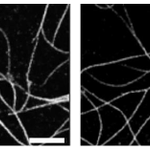



Gen-AI producing fancy data augmentation for Super-resolution microscopy! Great work @AlonSaguy and team! @HenriquesLab @HeilemannLab

If robots could dream of microtubules, how would they look like? An amazing story by Alon Saguy, @ShechtmanLab and colleagues. Proud we could contribute to it
onlinelibrary.wiley.com/doi/…


Great work @yevgenin @DanielleSapir et al.!

Thrilled to share OM2Seq! We applied #AI methods to #Genomics, improving the speed and accuracy of optical genome mapping for fast diagnostics of diseases. @DanielleSapir, Tahir Detinis Zur, Nir Weinberger, @boknilev, @hagenom, @ShechtmanLab
🔬🚀
We used the Incucyte system by @SartoriusGlobal. All this and more in our latest paper: link.springer.com/article/10…. Big thanks and congrats to all co-authors and collaborators: Nadav, @EliasNehme9 Noam, Ilana, Reut, Boris, Paul, @AlaloufOnit , @SartoriusGlobal !
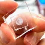
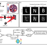
This enables PSF engineering in space-limited microscopy systems, e.g. that fit into incubators. Uses include extended depth-of-field and snapshot 3D spheroid from 2D, using algorithms by @EliasNehme9. Bonus: Obtaining training images is easy with a high-throughput microscope.
PSF engineering typically requires a 4-f system, but this is not always feasible, e.g. due to size constraints. Nadav Opatovski attached a phase mask directly to the objective lens, see beads defocusing here. Tetrapod mask shown here (wait for the end):
Thanks!
Re simultaneous processes - there will be a complexity-performance tradeoff that would be interesting to check (TBD). Of course if the processes are labeled orthogonally (e.g. different colors) then each one will just be solved separately.
🥳 DBlink is now peer reviewed and improved! Online in @naturemethods: rdcu.be/dhUmZ
Congrats @AlonSaguy, @AlaloufOnit, @HeilemannLab et al.!

New prepint just out! Live cell single molecule localization microscopy at >3 fps. The information is mostly there - just spread out in space and time :)
We use a recurrent neural net (LSTM) + some assumptions to extract it. All details here:
biorxiv.org/content/10.1101/…
More info in the preprint: biorxiv.org/content/10.1101/………, where we demonstrate the use-case of generating training data for CARE (by
@martweig et al.)
3/4
This is a very ongoing project, with many open questions: what are the performance limits? How much can we really gain beyond the information in the training data? and of course: what would be the best uses of something like this in super-resolution microscopy?
4/4
Alon Saguy et al. trained a diffusion model to generate synthetic images of super-resolution microscopy images using data of only a few images from ShareLoc (shareloc.xyz).
2/4
One of these super-resolution microtubule images was captured by a microscope and the other one was generated by a diffusion model. Can you tell which is which? Try more here: clever-microscopy.com/this-m…
1/4

Interesting. Do you also get blinking-pairs mistaken for a vertically-elongated PSF (high Z)? I would expect to see a similar number of those.
Great work by @yevgenin applying information theory to optical genome mapping, with implications to optimal label design
New #preprint!
TLDR: Applying #informationtheory on #OpticalGenomeMapping🧬, one can choose pattern-labelling #enzymes giving #10x accuracy! 🤯
biorxiv.org/content/10.1101/…
To illustrate, let’s play a game! ⬇️ (1/4)
