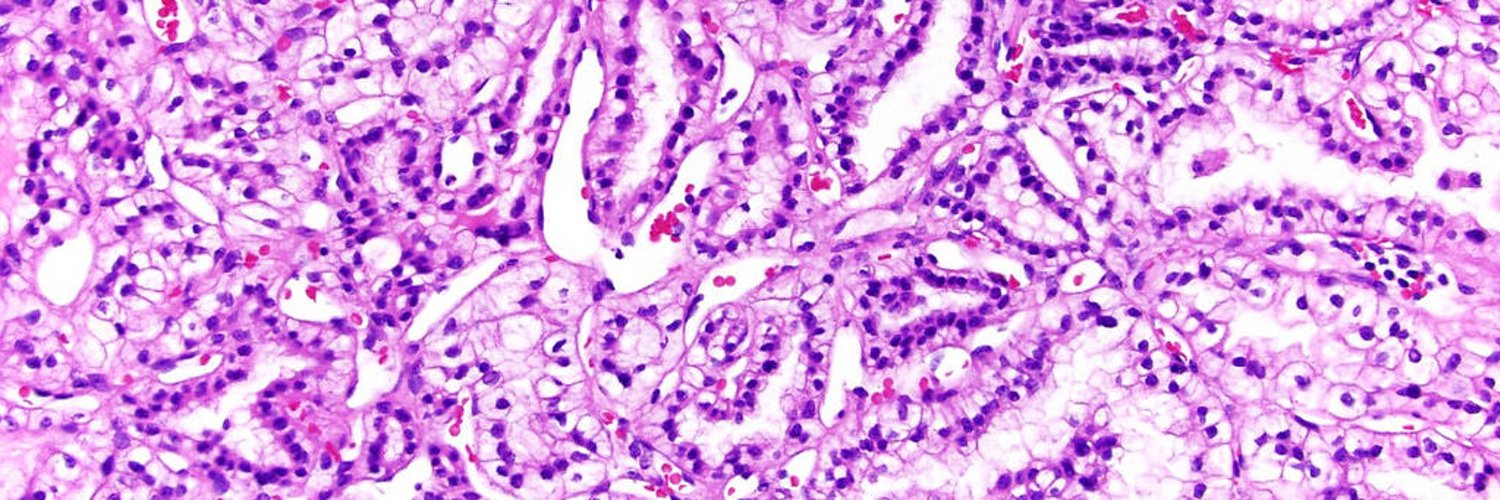

https://xitter.cf/t.co/5BLtUpcmNF is a free, no-registration website with a unique, comprehensive, regularly updated textbook of surgical and clinical pathology, and more!
Joined July 2009
- Tweets3K
- Following5.2K
- Followers25.7K
- Likes1.4K






PathologyOutlines.com retweeted
Stay in the loop with PathologyOutlines.com!
»facebook.com/pathologyoutlin…
»instagram.com/pathoutlines
»threads.net/@pathoutlines
»linkedin.com/company/patholo…
»reddit.com/r/PathologyOutlin…
»bsky.app/profile/pathologyou…
»youtube.com/c/PathologyOutli…
»pathologyoutlinesblog.wordpr…
#PathologyOutlines #PathTwitter

PathologyOutlines.com retweeted

#slidearchiveseries metanephric adenoma #gupath #pathtwitter #pathoutpic
🔵 benign, blue tumor, tightly packed tubules/papillary structures, frequent calcifications
🔵 PAX8/WT1/CD57/BRAF (+)
🔵 DDx: 👨🏻 PRCC, solid variant and 👦🏼 epithelial predominant Wilms tumor


PathologyOutlines.com retweeted
#slidearchiveseries mucinous tubular and spindle cell carcinoma #gupath #pathtwitter #pathoutpic
🔬 tubular and spindle cell growth patterns associated myxoid stroma
🎨 CK7, AMACR, VSTM2A (PMID 30285995) @drmehrarohit
🧬 No trisomies 7 / 17 or loss of Chr Y


PathologyOutlines.com retweeted
#slidearchiveseries papillary renal neoplasm with reverse polarity #gupath #pathtwitter #pathoutpic
🕵🏻♀️ usually asymptomatic, incidentally found, clinically indolent
🔬 thin papillary/tubulopapillary growth, apically located, low grade nuclei
🎨 CK7, GATA3, L1CAM
🧬 KRAS mutation


PathologyOutlines.com retweeted

F/60+, ileocecal recection.
Low grade appendiceal mucinous neoplasm (LAMN) .
Dilated appendix with perforation and diverticula, with hemorrhages, with luminal and extra-appendiceal mucin.
1/3🧵🔽
#Grosspath #pathology #Gipath #pathoutpic

ALT Red/white arrow in the projection of the base/entrance of the appendix.

PathologyOutlines.com retweeted
#slidearchiveseries chromophobe renal cell carcinoma #gupath #pathtwitter #pathoutpic
✅ solid sheet-like architecture
✅ plant-like cell borders (vegetable cells)
✅ irregular, wrinkled nuclei (raisinoid)
✅ binucleation and perinuclear halos
🎨 CK7, CD117, Hale colloidal iron


PathologyOutlines.com retweeted
#macrovsmicro partial nephrectomy, variegated red-brown to yellow-tan, focally calcified mass #gupath #pathtwitter #pathoutpic #kidneytumor

PathologyOutlines.com retweeted
🎯 metastatic renal medullary carcinoma
🔬reticular, cribriform, and cord-like patterns; intratumoral inflammatory infiltrate (sometimes abscess forming); fibrotic stroma with myxoid areas
✨ metastasis common to lung, liver, LN, adrenal gland, RP, etc #pathoutpic


PathologyOutlines.com retweeted

Granular cell tumor can occur anywhere in the digestive tract. Esophagus is most common, but here's a large case from the colon. Classic histology, but getting confirmatory IHC (positive S100 & CD68) never hurts. shorturl.at/cdglN #pathology #gipath #PathTwitter #PathOutPic


PathologyOutlines.com retweeted
Benign polyp-forming lesion, or Harry Potter spell? I give you ... gastritis cystica polyposa! pathologyoutlines.com/topic/… #pathology #gipath #PathTwitter #PathOutPic


PathologyOutlines.com retweeted
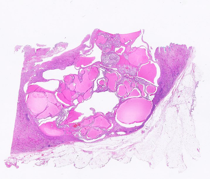
#slidearchiveseries clear cell papillary renal cell tumor #gupath #pathtwitter #pathoutpic
🫘 pT1 tumor (G1 or G2) (>95%)
🔬 mix of tubules, cysts, papillae, and small nests, in a fibromyomatous stroma
🎹 polarized nuclei “piano key-like”
🎨 CK7, CAIX “cup-like” basolateral


PathologyOutlines.com retweeted
#slidearchiveseries papillary renal cell carcinoma #gupath #pathtwitter #pathoutpic
🔬 small cuboidal cells arranged in a single layer around papillary cores containing nests of foamy macrophages
🧬 chr 7 and 17 gains, loss of Y; MET gene mutations {PMID: 26536169, 10433944}


PathologyOutlines.com retweeted
#slidearchiveseries urothelial carcinoma, micropapillary subtype
#gupath #pathtwitter #pathoutpic
🔬 small tumor cell clusters without fibrovascular cores, floating in lacunar spaces
⚠️ may resemble vascular invasion


PathologyOutlines.com retweeted
#slidearchiveseries amyloidosis of seminal vesicle #gupath #pathtwitter #pathoutpic
🧓🏻 localized, age-related finding, not typically related to a systemic process
🎨 Congo Red, pink-red staining pattern on light microscopy 🍏 apple-green birefringence on polarization


PathologyOutlines.com retweeted
#slidearchiveseries intratubular large cell hyalinizing Sertoli cell neoplasia
#gupath #pathtwitter #pathoutpic
💡frequent in Peutz-Jeghers syndrome
💡estrogen effects (gynecomastia, adv bone age)
💡conservative mgmt with aromatase inhibitors
📄 pubmed.ncbi.nlm.nih.gov/1752…


PathologyOutlines.com retweeted
M/30+, orchidectomy, testicular atrophy.
Dense fibrovascular tissue, foci of calcification and deposits of hemosiderin, cholesterol clefts.
#grosspath #GUPath #testis #pathology #pathoutpic

ALT Atrophy developed after hernioplasty for an inguinal hernia. In the top photo,for comparison,there is another case where a normal testicle was removed for a reason unrelated to the pathology in it.


PathologyOutlines.com retweeted
Granulomatous appendicitis can bring up a wide differential diagnosis. Don't forget interval appendicitis, given that interval appendectomies are becoming much more common. This case is one such example. tinyurl.com/2ndx69rz / #pathology #PathTwitter #gipath #PathOutPic


PathologyOutlines.com retweeted
#slidearchiveseries scrotal calcinosis
#gupath #dermpath #pathoutpic
🔬intradermal nodules varying in size and number, foreign body giant cell reaction in surrounding tissue
📊 differentials include calcification of epidermal or pilar cyst
🤔 idiopathic or dystrophic


PathologyOutlines.com retweeted
Incidental pelvic lymph node #lymphangioleiomyomatosis (LAM)
✴️clinically occult lymph node LAM can be detected during surgical staging of pelvic cancer; not commonly linked to pulmonary LAM/TSC; pts should be evaluated for both (PMID 25786086)
#gynpath #pathtwitter #pathoutpic


