

Professionally designed high-yield medical notes to help you prepare for exams and excel in clinical practice📝 - visit our website for e-books ⬇️
Joined September 2015
- Tweets2.4K
- Following6
- Followers7.2K
- Likes214




Pinned Tweet
MedicoNotes is an online medical education platform that provides professionally designed high-yield medical notes to help doctors prepare for exams and excel in medical practice.
💡 Study smarter with our medical notes and improve your exams results!
👉 A MUST-HAVE for all Medical, MBBS, MD, & MBChB students, PA, nursing students, as well as professional exams including USMLE, PLAB and UK Royal College exams.
👉 ALL Notes are delivered instantly in print-ready PDF format so you can study the way you learn best!
👉Free sample downloads are available at our website: mediconotes.com
———————-
#medstudentnotes #medstudent #medicalstudent #MDstudent #studentdoctor #medicine #medicalschool #medschool #medicalnotes #doctors #premed #futuredoctors #usmleprep #usmle #medstudentlife #medstudy #mednotes #medicalstudy #medicalnotes #medico #PAstudent #MDlife #mbchb
🔄 Renin-Angiotensin-Aldosterone System (RAAS)
📊 Mnemonic: “RAAS Effects”
Retain Na⁺
Aldosterone ↑
ADH released
Sympathetic activated
📚 Visit our website for more high-yield medical notes: mediconotes.com

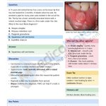
🩺 MEDICAL MCQ: NON-HEALING LOWER LIP ULCER
A 71-year-old retired farmer presents with a lower lip ulcer that has persisted for 2 months and bleeds when eating.
Remember the 3 S’s:
☀️ Sun exposure
🚬 Smoke exposure
🐌 Slow-healing sore
🌐 mediconotes.com

💡NEWLY LAUNCHED BOOK!
📚Complete Differential Diagnosis Collection
Master clinical reasoning with the complete MedicoNotes Differential Diagnosis.
—————-
🔗 Visit our website to download the book NOW:
mediconotes.com
——————
#Medicine #Doctors #USMLE #meded


🧬 MEN Syndromes — Distinguish Them in 60 Seconds
⸻
1️⃣ MEN 1 — Wermer Syndrome
The “3 Ps”
🔸 Pituitary Tumour → e.g., Prolactinoma
🔸 Parathyroid Hyperplasia → most common initial presentation → hypercalcaemia
🔸 Pancreatic Islet Cell Tumours → Gastrinoma (Zollinger–Ellison) + Insulinoma
🧬 Gene: MEN1 (Chromosome 11) → Tumour suppressor mutation
⸻
2️⃣ MEN 2A — Sipple Syndrome
The “2 Ps + 1 M”
🔸 Parathyroid Hyperplasia → hypercalcaemia
🔸 Phaeochromocytoma → adrenal gland tumour
🔸 Medullary Thyroid Carcinoma (MTC)
🧬 Gene: RET proto-oncogene (Chromosome 10) → Activating mutation
⸻
3️⃣ MEN 2B — Mucosal & Marfanoid
The “1 P + 2 Ms”
🔸 Phaeochromocytoma
🔸 Medullary Thyroid Carcinoma → most aggressive form, early onset
🔸 Mucosal Neuromas + Marfanoid Habitus
⚠️ NO parathyroid disease — key distinguishing feature!
🧬 Gene: RET proto-oncogene (Chromosome 10) → Activating mutation
⸻
4️⃣ The Master Comparison
➊ MEN 1 → Pituitary + Parathyroid + Pancreas (MEN1 gene, Chr 11)
➋ MEN 2A → MTC + Pheo + Parathyroid (RET gene, Chr 10)
➌ MEN 2B → MTC + Pheo + Neuromas/Marfanoid — NO parathyroid (RET gene, Chr 10)
⸻
🧩 Exam Pearls
🔹 MEN 2B is the MOST aggressive — presents earliest with MTC
🔹 Phaeochromocytoma rule → ALWAYS give alpha-blocker BEFORE beta-blocker to prevent hypertensive crisis
🔹 MEN 1 vs MEN 2 genetics → Tumour suppressor (MEN1) vs Activating oncogene (RET)
🔹 Hypercalcaemia in MEN → think Parathyroid (MEN 1 and 2A only — NOT 2B)
⸻
📚 Master MEN syndromes and all of endocrinology in our high-yield endocrinology book:
🔗 mediconotes.com
⸻
#MENSyndrome #usmle #Endocrinology #MedicalEducation #doctor

🌀 7 CAUSES OF VERTIGO
Vertigo is a symptom, not a diagnosis.
When a patient complains of “dizziness,” the key question is:
❓ Is this true vertigo (a false sensation of movement) or something else?
Correctly distinguishing peripheral from central causes is essential, as some causes are benign, while others may represent a neurological emergency.
🔑 Common Causes of Vertigo
1️⃣ BPPV (Benign Paroxysmal Positional Vertigo)
• Most common cause
• Brief episodes triggered by head movement
• No hearing loss
• Positive Dix-Hallpike test
• Treated with the Epley manoeuvre
2️⃣ Vestibular Neuritis
• Acute severe vertigo lasting days
• Often follows a viral illness
• Nausea and vomiting common
• No hearing loss
3️⃣ Ménière Disease
• Recurrent episodes of vertigo
• Fluctuating hearing loss
• Tinnitus and aural fullness
• Usually unilateral
4️⃣ Vestibular Migraine
• Vertigo associated with migraine features
• Photophobia, phonophobia, aura
• May occur without headache
5️⃣ Labyrinthitis
• Vertigo + hearing loss
• Often post-viral
• Auditory and vestibular symptoms occur together
6️⃣ Acoustic Neuroma (Vestibular Schwannoma)
• Progressive unilateral hearing loss
• Tinnitus
• Gradual onset imbalance or vertigo
7️⃣ Central Causes 🚨
• Stroke
• Multiple sclerosis
• Cerebellar tumours
• Brainstem lesions
⚠️ Red Flags Suggesting a Central Cause
🚨 Diplopia
🚨 Dysarthria
🚨 Limb weakness
🚨 Severe ataxia
🚨 New headache
🚨 Persistent neurological deficits
🩺 Clinical Approach
✔️ Take a careful history
✔️ Examine for nystagmus
✔️ Perform the HINTS examination when appropriate
✔️ Assess hearing
✔️ Look for focal neurological signs
💡 High-Yield Pearl
Peripheral vertigo is common, but never miss a central cause.
Remember:
🧠 Acute vertigo + neurological signs = Stroke until proven otherwise.
📚 Save this post and visit our website for innovative high-yield medical notes:
MedicoNotes.com
#Vertigo #Neurology #BPPV #MeniereDisease #vestibularneuritis

💡 Guide to Hypothyroidism Management
⸻
1️⃣ Clinical Features
🔹 Common Symptoms
➊ Cold intolerance
➋ Fatigue
➌ Weight gain
➍ Constipation
➎ Bradycardia
➏ Mental slowing
📌 Suspect hypothyroidism in patients with metabolic slowing and fatigue
⸻
2️⃣ Diagnosis — TSH is the Gold Standard
🧪 Primary Hypothyroidism
⬆️ TSH + ⬇️ Free T4
🧪 Subclinical Hypothyroidism
⬆️ TSH + Normal Free T4
🧪 Central Hypothyroidism
⬇️/Normal TSH + ⬇️ Free T4
⚠️ Central causes suggest pituitary/hypothalamic disease
⸻
3️⃣ Investigating the Cause
🔬 Common Tests
➊ Anti-TPO antibodies
➋ Anti-thyroglobulin antibodies
📌 Hashimoto thyroiditis is the most common cause
🧠 In suspected central disease, assess:
• ACTH/cortisol
• Prolactin
• LH/FSH & GH
⸻
4️⃣ Treatment of Choice
💊 Levothyroxine (T4)
📌 Initial Dosing
➊ Healthy adults → ~1.6 mcg/kg/day
➋ Elderly/CAD → Start low (12.5–25 mcg/day)
➌ Pregnancy → Increase dose by 30–50%
⸻
5️⃣ Important Dosing Tips
⏰ Take on an empty stomach
🚫 Separate from:
• Iron
• Calcium
• Antacids
📌 Recheck TSH every 6 weeks after dose changes
⸻
6️⃣ Subclinical Hypothyroidism
✅ Usually treat if:
• TSH >10 mIU/L
• Symptomatic
• Positive antibodies
• Pregnancy/fertility concerns
⸻
7️⃣ Myxedema Coma 🚨
⚠️ Features
• Hypothermia
• Bradycardia
• Hypotension
• Altered mental status
🚑 Management
➊ Airway/supportive care
➋ Warm IV fluids
➌ IV levothyroxine
➍ IV hydrocortisone
⸻
💡 Clinical Pearls
➊ High TSH + low FT4 = overt hypothyroidism
➋ Low/normal TSH + low FT4 = central hypothyroidism
➌ Exclude adrenal insufficiency before high-dose thyroid replacement
➍ Pregnancy increases levothyroxine requirements
⸻
📚 Visit our website to download our new Endocrinology Book:
🌐 mediconotes.com
#Endocrinology #Hypothyroidism #MedicalEducation #USMLE #meded

🧠 Stroke Localization Made Simple
⸻
1️⃣ MCA Stroke (Middle Cerebral Artery)
➊ Face & arm weakness > leg weakness
➋ Contralateral sensory loss
➌ Dominant side → Aphasia
➍ Non-dominant side → Neglect
💡 Remember:
MCA = “M” for Mouth & aRM involvement
⸻
2️⃣ ACA Stroke (Anterior Cerebral Artery)
➊ Leg weakness > arm weakness
➋ Personality changes
➌ Abulia (loss of motivation)
➍ Urinary incontinence
💡 Think:
ACA affects the medial brain → lower limb area
⸻
3️⃣ PCA Stroke (Posterior Cerebral Artery)
➊ Contralateral homonymous hemianopia
➋ Visual symptoms
➌ Alexia without agraphia
➍ Macular sparing
💡 Classic clue:
“I can write… but I can’t read”
⸻
4️⃣ Lateral Medullary Syndrome (PICA)
➊ Dysphagia & hoarseness
➋ Vertigo, nystagmus, ataxia
➌ Ipsilateral facial sensory loss
➍ Contralateral body pain/temperature loss
➎ Horner syndrome
💡 Crossed findings = brainstem lesion
⸻
5️⃣ Medial Medullary Syndrome (ASA)
Remember the “3 M’s”:
➊ Medial lemniscus → ↓ vibration/proprioception
➋ Motor pathway → contralateral weakness
➌ Medulla CN XII → tongue deviation
⸻
🎯 Stroke localization becomes easy when you recognize the pattern!
📚 Master neurology with our high-yield Neurology Book 🧠
Available now at:
🌐 mediconotes.com
#Neurology #Stroke #MedEd #USMLE #Medical

💡HYPOXIA: LOW O₂, MANY CAUSES!
Hypoxia is a sign, not a diagnosis. Understanding the mechanism helps you identify the cause and treat it quickly.
⸻
1️⃣ V/Q Mismatch
📍 Most common cause of hypoxia
🔹 Ventilation and perfusion are mismatched
🔹 Seen in asthma, COPD, pneumonia
💎 Pearl:
Improves with supplemental oxygen.
⸻
2️⃣ Shunt
📍 Blood bypasses ventilated alveoli
🔹 No gas exchange occurs
🔹 Seen in ARDS, severe pneumonia, atelectasis
💎 Pearl:
Does NOT significantly improve with oxygen.
⸻
3️⃣ Diffusion Defect
📍 Impaired gas transfer across alveolar membrane
🔹 Pulmonary fibrosis
🔹 Interstitial lung disease
💎 Pearl:
Worse during exercise.
⸻
4️⃣ Hypoventilation
📍 Reduced alveolar ventilation
🔹 CNS depression
🔹 Neuromuscular disorders
🔹 Obesity hypoventilation
💎 Pearl:
↑ PaCO₂ + ↓ PaO₂
⸻
5️⃣ Low FiO₂
📍 Reduced inspired oxygen
🔹 High altitude
🔹 Poorly ventilated environments
💎 Pearl:
Improves with increased FiO₂.
⸻
6️⃣ Pulmonary Embolism (PE)
📍 Perfusion defect
🔹 Sudden dyspnea
🔹 Pleuritic chest pain
🔹 Tachycardia
💎 Pearl:
Ventilation present, perfusion absent.
⸻
7️⃣ ARDS
📍 Diffuse alveolar injury
🔹 Severe hypoxemia
🔹 Pulmonary edema
🔹 Reduced lung compliance
💎 Pearl:
Refractory hypoxemia despite oxygen therapy.
⸻
💎 High-Yield Exam Pearls
🫁 V/Q Mismatch → Most common
🫁 Shunt → Doesn’t improve with O₂
🫁 Diffusion Defect → Worse on exertion
🫁 Hypoventilation → High CO₂
🫁 Low FiO₂ → High altitude
🫁 PE → Ventilation without perfusion
🫁 ARDS → Refractory hypoxemia
⸻
📚 Master Respiratory Medicine the High-Yield Way with the MedicoNotes Respiratory Book.
🌐 Visit our website: mediconotes.com
#RespiratoryMedicine #Hypoxia #MRCP #USMLE #PLAB

🧠 8 Movement Disorder Localizations
Movement disorders can arise from lesions at multiple levels of the nervous system. Recognizing the clinical pattern is key to accurate localization and diagnosis.
1️⃣ Basal Ganglia
🔹 Disorders: Parkinsonism, chorea, dystonia
🔹 Clues: Resting tremor, rigidity, bradykinesia, abnormal postures
🔹 Common causes: Parkinson’s disease, Huntington’s disease, drugs, stroke
2️⃣ Thalamus
🔹 Disorders: Tremor, hemiballismus
🔹 Clues: Contralateral involuntary movements
🔹 Common causes: Stroke, hemorrhage, multiple sclerosis
3️⃣ Cerebellum
🔹 Disorders: Ataxia, intention tremor, dysmetria
🔹 Clues: Gait instability, nystagmus, dysdiadochokinesia
🔹 Common causes: Stroke, tumors, alcohol, multiple sclerosis
4️⃣ Brainstem
🔹 Disorders: Tremor, dystonia, parkinsonism
🔹 Clues: Cranial nerve deficits with long tract signs
🔹 Common causes: Stroke, tumors, neurodegenerative diseases
5️⃣ Spinal Cord
🔹 Disorders: Spasticity, clonus, segmental myoclonus
🔹 Clues: UMN signs below the lesion with sensory level
🔹 Common causes: Trauma, transverse myelitis, multiple sclerosis
6️⃣ Motor Cortex
🔹 Disorders: Focal motor seizures, apraxia, myoclonus
🔹 Clues: Cortical signs, seizures, contralateral weakness
🔹 Common causes: Stroke, tumors, encephalitis
7️⃣ Peripheral Nerves
🔹 Disorders: Fasciculations, weakness, occasional tremor
🔹 Clues: LMN signs including atrophy and fasciculations
🔹 Common causes: Peripheral neuropathy, entrapment, motor neuron disease
8️⃣ Neuromuscular Junction / Muscle
🔹 Disorders: Fatigable weakness, myopathies
🔹 Clues: Ptosis, diplopia, proximal weakness, fatigability
🔹 Common causes: Myasthenia gravis, inflammatory myopathies, muscular dystrophies
————————
📚 Master neurological localization with our comprehensive MedicoNotes Neurology Book—packed with high-yield diagrams, clinical pearls, and exam-focused summaries.
🌐 mediconotes.com
————————-
#Neurology #MovementDisorders #ParkinsonDisease #NeurologyResident #MedicalEducation

❤️ CARDIOLOGY EMERGENCY CRASH CARDS
The difference between life and death in cardiology is often measured in minutes.
⸻
1️⃣ STEMI Crash Card
➊ Recognize chest pain + ST elevation
➋ Activate reperfusion pathway immediately
➌ Door-to-balloon < 90 minutes
➍ Time = Myocardium ❤️
⸻
2️⃣ Arrhythmia Crash Card
➊ SVT → Adenosine
➋ AF with RVR → Rate control
➌ VT with pulse → Amiodarone/Cardioversion
➍ VF/Pulseless VT → Defibrillate NOW ⚡
⸻
3️⃣ Acute Heart Failure
➊ Oxygen + sit upright
➋ Nitrates if appropriate
➌ IV diuretics
➍ Escalate early if respiratory distress
⸻
4️⃣ Cardiogenic Shock
➊ Identify poor perfusion early
➋ Monitor lactate & urine output
➌ Vasopressors + inotropes when needed
➍ Urgent revascularization saves lives
⸻
5️⃣ Night Duty Survival Card
➊ ECG recognition made simple
➋ Emergency drug doses
➌ ACLS essentials
➍ Quick-reference bedside guide
⸻
💡 High-Yield Exam Pearl:
STEMI → Reperfuse
SVT → Adenosine
AF → Rate Control
VF/VT → Defibrillate
Shock → Restore Perfusion
⸻
📚 Master Cardiology with the MedicoNotes Cardiology Book
🌐 mediconotes.com
#Cardiology #ECG #STEMI #MedicalEducation #MedicoNotes
🤖 Made with AI


🧠 7 CAUSES OF ATAXIA
Ataxia is a sign, not a diagnosis. The key is to localize the lesion first, then identify the cause.
⸻
1️⃣ Cerebellar Ataxia
📍 Lesion in the cerebellum or its pathways
🔹 Dysmetria
🔹 Intention tremor
🔹 Nystagmus
🔹 Scanning speech
🔹 Broad-based gait
🧠 Causes:
➊ Stroke
➋ Tumor
➌ Cerebellitis
➍ Degenerative disease
⸻
2️⃣ Sensory Ataxia
📍 Loss of proprioception from dorsal columns or peripheral nerves
🔹 Positive Romberg sign
🔹 Stamping gait
🔹 Worse in darkness
🔹 Loss of vibration sense
🧠 Causes:
➊ Vitamin B12 deficiency
➋ Tabes dorsalis
➌ Peripheral neuropathy
➍ Friedreich ataxia
⸻
3️⃣ Vestibular Ataxia
📍 Vestibular apparatus or vestibular nerve lesion
🔹 Vertigo
🔹 Nausea & vomiting
🔹 Nystagmus
🔹 Imbalance
🔹 Falls toward affected side
🧠 Causes:
➊ Vestibular neuritis
➋ Labyrinthitis
➌ Ménière disease
➍ Vestibular schwannoma
⸻
4️⃣ Alcohol-Related Ataxia
🍺 Chronic alcohol toxicity
🔹 Wide-based gait
🔹 Cerebellar degeneration
🔹 Poor coordination
🔹 Nutritional deficiency
🧠 Associated with:
➊ Thiamine deficiency
➋ Wernicke encephalopathy
———
5️⃣ Vitamin B12 Deficiency Ataxia
🟠 Subacute Combined Degeneration
🔹 Sensory ataxia
🔹 Positive Romberg
🔹 Paresthesia
🔹 Loss of vibration sense
🔹 Cognitive symptoms
🧠 Examination:
➊ Reduced proprioception
➋ Dorsal column signs
➌ Spasticity (late)
⸻
6️⃣ Multiple Sclerosis (MS)
🧠 Demyelination affecting cerebellar pathways
🔹 Ataxia
🔹 Diplopia
🔹 Optic neuritis
🔹 Limb weakness
🔹 Sensory symptoms
⸻
7️⃣ Cerebrovascular Disease (Stroke)
🚨 Acute onset cerebellar or brainstem lesion
🔹 Sudden ataxia
🔹 Vertigo
🔹 Vomiting
🔹 Dysarthria
🔹 Cranial nerve deficits
🧠 Causes:
➊ Cerebellar infarction
➋ Cerebellar hemorrhage
➌ Brainstem stroke
⸻
📚 Master Neurology the High-Yield Way with the MedicoNotes Neurology Book.
🌐 Visit our website: mediconotes.com
#Neurology #Ataxia #MRCP #USMLE #plab

❤️ HFpEF Management — 2026 ACC Update
HFpEF management is becoming more phenotype-driven, with newer therapies targeting cardiovascular, renal and metabolic risk—not just congestion.
1️⃣ Confirm the Diagnosis
Think HFpEF in patients with dyspnoea, oedema or exercise intolerance.
Use clinical assessment, echo, natriuretic peptides and scores such as H₂FPEF / HFA-PEFF when helpful.
Always consider mimics and alternative causes of breathlessness.
2️⃣ SGLT2 Inhibitors — Core Therapy
Empagliflozin or dapagliflozin should be considered broadly in HFpEF, regardless of diabetes status.
They reduce HF events and provide important cardiovascular–renal benefits.
3️⃣ Consider Finerenone
The 2026 pathway incorporates evidence supporting non-steroidal MRA therapy in HFpEF.
Check renal function and potassium before and during treatment.
4️⃣ Treat the Phenotype
Obesity: weight reduction, exercise and incretin-based therapy where appropriate.
Hypertension: optimise BP control.
AF, CAD, CKD and diabetes: actively identify and manage associated disease.
ARNI/MRA/ARB therapy may be considered in selected patients rather than used uniformly in everyone.
5️⃣ Don’t Forget Symptoms
Use diuretics for congestion.
Encourage regular exercise, weight optimisation and management of sleep apnoea where relevant.
Avoid unnecessary medications that worsen chronotropic incompetence or exercise tolerance.
💡 High-Yield Pearl:
HFpEF is not one disease phenotype. The modern approach is:
Confirm HFpEF → SGLT2i → manage congestion → treat comorbidities → tailor therapy to phenotype.
📚 Explore our MedicoNotes Cardiology Book for high-yield cardiology concepts, treatment algorithms and exam-focused revision:
🌐 mediconotes.com
#HFpEF #HeartFailure #Cardiology #MedicalEducation #MedicoNotes
🤖 Made with AI


🧠 MCA vs ACA vs PCA — Localise the Stroke in 5 seconds!
⸻
1️⃣ MCA — Middle Cerebral Artery
🔸 Contralateral Face & Arm weakness
🔸 Contralateral Face & Arm sensory loss
🔸 Aphasia → if dominant hemisphere
🔸 Gaze deviation TOWARD the lesion
🔸 Visual field: Contralateral Homonymous Hemianopia
🧩 Why? MCA supplies the lateral cortex — face, arm, and language areas
⸻
2️⃣ ACA — Anterior Cerebral Artery
🔸 Contralateral LEG weakness (arm spared!)
🔸 Contralateral LEG sensory loss
🔸 Abulia / Apathy → behavioural changes
🔸 Urinary incontinence
🔸 Visual field: Contralateral Homonymous Hemianopia
🧩 Why? ACA supplies the medial frontal and parietal lobes — leg area and motivation circuits
⸻
3️⃣ PCA — Posterior Cerebral Artery
🔸 Contralateral Visual Field Loss → often isolated
🔸 Visual Hallucinations → Charles Bonnet Syndrome
🔸 Visual Agnosia → impaired visual recognition
🔸 Visual field: Contralateral Homonymous Hemianopia with macular sparing
🧩 Why? PCA supplies the occipital lobe (vision) and inferior temporal lobe (visual memory)
⸻
🧩 The 5-Second Localisation Rule
➊ Face + Arm weak + Aphasia → MCA
➋ Leg weak + Behavioural change → ACA
➌ Vision loss only + Hallucinations → PCA
⸻
⚠️ The Exam Trap
🔹 All three can cause Contralateral Homonymous Hemianopia
🔹 ACA and MCA → full field defect
🔹 PCA → macular SPARING (occipital cortex has dual blood supply)
⸻
📚 Master all neurology conditions in our high-yield neurology book:
🔗 mediconotes.com
⸻
#StrokeSyndromes #Neurology #medicine #internalmedicine #usmle

💡 5 ELECTROLYTE EMERGENCIES YOU MUST NEVER MISS
⸻
1️⃣ Hyperkalemia (↑ K⁺)
➊ Clinical Features
• Muscle weakness
• Palpitations
• Life-threatening arrhythmias
➋ ECG Changes
• Peaked T waves
• PR prolongation
• Wide QRS
• Sine-wave pattern (pre-terminal)
➌ Emergency Treatment
• IV Calcium Gluconate → Stabilize myocardium
• Insulin + Dextrose → Shift K⁺ intracellularly
• Salbutamol
• Dialysis if severe
💎 Pearl:
Calcium protects the heart but does NOT lower potassium.
⸻
2️⃣ Hypokalemia (↓ K⁺)
➊ Causes
• Vomiting
• Diarrhea
• Loop/Thiazide diuretics
• Insulin excess
➋ ECG Changes
• Flattened T waves
• Prominent U waves
➌ Treatment
• Oral or IV potassium replacement
💎 Pearl:
Refractory hypokalemia? Always check magnesium.
⸻
3️⃣ Severe Hyponatremia (↓ Na⁺)
➊ Clinical Features
• Confusion
• Seizures
• Coma
➋ Causes
• SIADH
• Excess water intake
• Heart failure
• Cirrhosis
➌ Emergency Treatment
• 3% Hypertonic Saline for severe symptoms
⚠️ Critical Pearl:
Correct slowly to avoid Osmotic Demyelination Syndrome (ODS).
⸻
4️⃣ Hypercalcemia Crisis (↑ Ca²⁺)
➊ Classic Presentation
🦴 Bones
🪨 Stones
🤢 Groans
🧠 Psychiatric overtones
➋ ECG Changes
• Short QT interval
➌ Treatment
• IV Normal Saline
• Calcitonin
• Bisphosphonates
• Dialysis if severe
💎 Pearl:
The two most common causes are:
• Primary Hyperparathyroidism
• Malignancy
⸻
5️⃣ Hypocalcemia & Hypomagnesemia
➊ Clinical Features
• Tetany
• Seizures
• Perioral numbness
• Chvostek sign
• Trousseau sign
➋ ECG Changes
• Long QT interval
• Torsades de Pointes (low Mg²⁺)
➌ Treatment
• IV Calcium Gluconate
• IV Magnesium Sulfate for torsades
💎 Pearl:
Alcohol misuse + low K⁺ + low Ca²⁺ = Check magnesium immediately.
⸻
⸻
📚 Master electrolyte disorders, acid-base balance, renal physiology, and emergency medicine with our comprehensive Nephrology Book.
🌐 Visit: mediconotes.com
#Nephrology #Electrolytes #Hyperkalemia #Hyponatremia #usmleprep

🧠 STROKE
Stroke occurs when blood flow to part of the brain is interrupted, causing sudden neurological dysfunction. Rapid recognition and treatment are essential to reduce permanent disability.
🔑 Classic Warning Signs
• Sudden facial droop
• Arm or leg weakness
• Speech difficulty
• Visual loss
• Severe imbalance or ataxia
• Sudden severe headache, especially in hemorrhagic stroke
💡 Remember FAST
F – Face drooping
A – Arm weakness
S – Speech difficulty
T – Time to seek emergency help
🧪 Diagnosis
• Urgent non-contrast CT head to exclude hemorrhage
• CT angiography if large-vessel occlusion is suspected
• MRI diffusion imaging is highly sensitive for acute ischemic stroke
• ECG, blood glucose and vascular risk assessment help identify the cause
💊 Management
Ischemic stroke:
• IV thrombolysis in eligible patients
• Mechanical thrombectomy for selected large-vessel occlusions
• Antiplatelet therapy when appropriate
Hemorrhagic stroke:
• Control blood pressure
• Reverse anticoagulation when required
• Urgent neurosurgical assessment in selected cases
⚠️ HIGH-YIELD PEARL:
Sudden focal neurological deficit should be treated as stroke until proven otherwise.
🧠 Remember:
Face weakness + Arm weakness + Speech disturbance = Think STROKE and act FAST.
📚 Want more high-yield neurology made simple? Get our MedicoNotes Neurology Book for concise, visual and exam-focused revision.
🌐 mediconotes.com
#Stroke #MedicalEducation #MRCP #medicaleducation #usmle usmle
🤖 Made with AI
💡PHEOCHROMOCYTOMA — A Rare but High-Yield Diagnosis!
Pheochromocytoma is a rare catecholamine-secreting tumour, most commonly arising from the adrenal medulla. Excess adrenaline and noradrenaline can cause dramatic, episodic symptoms.
🔑 Classic Triad
🧠 Headache
💦 Sweating
❤️ Palpitations
Patients may also develop episodic hypertension, tremor, anxiety and pallor.
🧪 Diagnosis
• Plasma free or urinary fractionated metanephrines
• CT/MRI to localise the tumour
• Consider genetic evaluation in appropriate patients
💊 Management
Definitive treatment is surgical resection, but careful pre-operative preparation is essential.
⚠️ HIGH-YIELD PEARL:
Always start α-blockade BEFORE β-blockade. Starting a β-blocker first may cause unopposed α-adrenergic stimulation and a dangerous hypertensive crisis.
🧠 Remember:
Headache + Sweating + Palpitations + Episodic Hypertension → Think Pheochromocytoma!
📚 Want more high-yield endocrinology made simple? Check out our MedicoNotes Endocrinology Book for concise, visual and exam-focused revision.
🌐 MedicoNotes.com
#Pheochromocytoma #Endocrinology #USMLE #medicaleducation #doctorsofinstagram
🤖 Made with AI