

Royal Adelaide Hospital and @COTR_Adelaide University of Adelaide
- Tweets176
- Following208
- Followers178
- Likes411


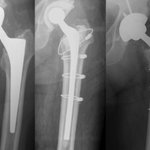








ALT Total hip arthroplasty (THA) for an acute acetabular fracture with a Trabecular Metal Revision Cup with multidirectional long intramedullary Screws (TMRCS) into the ilium, ischium, and pubis.


ALT The Bone & Joint Journal. Abstract text for article titled 'Estimation of two-year hospital costs of hip and knee periprosthetic joint infection treatments using activity-based costing'.


ALT Figure 3. (A) The RSA migration coordinate system for a right-sided tibia with X-axis pointing medially, Y-axis pointing superiorly, and Z-axis pointing anteriorly. (B) The CT migration coordinate system for right-sided tibia with X-axis pointing medially, Y-axis pointing posteriorly, and Z-axis pointing superiorly.


ALT Figure 1. RSA examinations showing the baseline (a) and 3 months (b) follow-up exami-nation of a patients. At 3 months, the anterolateral marker (red circle) in the PE insert is overprojected by the implant in the lateral radiograph. Using the consistent-marker method, only 4 of the 5 markers in the PE insert are selected at the 1-year follow-up (c) for the migration calculation (as these are the markers used at the 3 months examina-tion). With the all-marker method, all 5 available markers that can be matched to the reference examination are used for the migration calculation at the 1-year follow-up (d)

ALT Image depicts the centre of the cavity of the acetabular component estimated in 3D imaging.


ALT Bone & Joint Open Editor's Pick. Large articulations do not increase wear rates of thin second-generation highly cross-linked polyethylene liners at ten years. Callary et al. Citation: Bone Jt Open. 2023;4(11):839-845.

