






Pinned Tweet
A nasty met of melanoma in inguinal lymph node. The peritheliomatous distribution of melanoma cells (most are rhabdoid and plasmacytoid here) can be mistaken as perivascular formations of metastatic germ cell tumors without appropriate history. The patient has primary in big toe. – at Myanmar


Sections show mature pancreatic tissue composed of well-formed pancreatic acini, ducts, within the transverse mesocolon, consistent with pancreatic heterotopia. No cytologic atypia or malignancy is identified.
#pathology #GIPath #Pancreatic_heterotopia


Tracheal sampling on thyroid FNAC refers to accidental aspiration of tracheal mucosal cells during needle passage, especially when the thyroid nodule is close to the trachea. Smears may show benign ciliated columnar epithelial cells, mucus, and occasional squamous cells.

Skin biopsy shows a markedly dilated follicular infundibulum filled with laminated keratin and lined by acanthotic stratified squamous epithelium with radiating epithelial projections into the surrounding dermis, consistent with a dilated pore of Winer.
#pathology #dermpath


Rectal mucinous carcinoma with signet ring cell features in a young male.
#pathology #GIPath #mucinous_carcinoma #signets


Pitfall alert: This AE1/AE3 IHC from scalene nodes highlights scattered stromal/dendritic-appearing cells without convincing cytokeratin-positive metastatic epithelial deposits.
It may give rise to false positive interpretation of micrometastasis of carcinoma.
#pathology #IHCpath


Aggressive HER2-enriched micropapillary breast cancer.
Important point: in micropapillary carcinoma, strong basolateral membrane may still indicate HER2 positivity, even if it is not the classic complete circumferential staining pattern.
nature.com/articles/s41379-0…


Striking multivacuolated lipoblasts in a case of pleomorphic liposarcoma. Sometimes, extensive sampling is needed not to miss these lipoblasts as they are prerequisite for diagnosis of pleomorphic liposarcoma.
#pathology #BSTPath #Liposarcoma


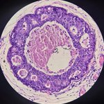
A focus of micropapillary DCIS characterized by ducts expanded by epithelial proliferation displaying micropapillary architecture. There is background invasive micropapillary carcinoma.
#pathology #breastpath #DCIS


Granulomatous inflammation affecting bone, marrow and joint capsule of right knee joint. The most likely cause for Myanmar's demographics is TB.
#pathology #BSTPath #Granuloma


A cute parathyoid incidentally found in right lobectomy specimen of thyroid done for multinodular goiter.
#pathology #endocrine #parathyroid


Cribriform DCIS with a rim of p63 positive myoepithelial cells surrounded by low grade invasive cribriform carcinoma of breast.
#pathology #IHC #Cribriform_carcinoma #breastpath

A pregnant lady with rapidly growing nodule in LSCS scar. A lovely example of scar deciduosis.
The diagnosis is straightforward on H&E sections.
#pathology #dermpath #Gynepath #deciduosis


