




Try something once, watch it fall over, file it under 'AI can't'. Six months later the task works. The grudge still doesn't.

A good lesson in AI is that you need to check back in on the latest with every 3 to 6 months to try something that wasn’t possible before and see if it works again.
At no point in AI’s history is that more true than today. Just the past six months of model progress around computer and tool use, less context rot, better reasoning, multimodality, and deeper domain expertise in a number of key fields, these models are vastly more capable on what they can execute.
This is opening up a new vector of what AI systems will be able to autonomously work on. Essentially, anything with an API (and even many things without), we can anticipate that an agent will be able to control and utilize as an underlying capability to complete a process.
This means that we are no longer dependent on *just* the models generating everything from whole cloth, but instead they can fully utilize existing systems (or new ones purpose built for them) very effectively. This creates immense leverage for agents.
Security agents can leverage existing data feeds and cyber tooling, IT agents that can execute workflows reliably across software stacks, data agents that can migrate data or processes between systems, architect and design agents can leverage CAD and design tools, and on and on. And any of today’s tools that don’t work will likely be replaced by agent-friendly or OSS tooling that does the same.
When you combine this with the domain understanding being trained into agents, you started to get autonomous collaborators for every job function and industry. This will open up a ton of new opportunities in AI if you build with this understanding in mind.
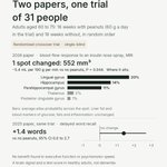
Nice to see this tested instead of argued about. Older vegetarians who lifted got stronger just like the meat eaters. A bit less muscle, but they also ate less protein, so that part isn't settled.

Older adults who lifted weights built muscle and strength on both a vegetarian and an omnivorous diet. The vegetarian group gained somewhat less muscle on average, but the trial could not tell whether that gap was real.
In the IMPACT randomized trial, 108 adults aged 55 and over (median age 66) did 12 weeks of resistance training, three sessions a week. Each was assigned to either a lacto-ovo vegetarian diet, with most protein from plants, or an omnivorous diet, with most protein from animal foods. Participants chose their own foods with dietary counseling and were given a daily protein target.
Both groups met that minimum target, but the vegetarian group ate about a fifth less protein overall. Arm and leg muscle, measured by DXA scan, increased by about 0.8 pounds in the vegetarian group and about 1.5 pounds in the omnivorous group. Leg press strength and chair-stand performance improved in both groups, with no clear difference between them.
The trial was designed to show whether the vegetarian diet was no worse for muscle gain, and it could not show that. The estimated difference was about 0.7 pounds in favor of the omnivorous diet, but the range of plausible results included no difference at all. The result is inconclusive, not proof that an omnivorous diet builds more muscle.
Because the vegetarian group also ate less protein, the effect of diet type and the effect of protein amount cannot be separated. The trial also lasted only 12 weeks.
What the study does show is that older vegetarians who train can build muscle and strength. Getting enough protein still matters, but this trial does not show that vegetarians cannot build muscle.
van Oppenraaij et al., American Journal of Clinical Nutrition, 2026.

Autonomous labs I can picture. "Master biology" I'll believe when one finds something a human lab missed.

Didn't know this one.
A deer's rumen is a fermentation vat. Dump corn on a winter browse diet and starch bacteria take over, acid spikes. The worst die in 24-72 hours.
Deer used to corn cope better. The sudden switch is the killer.

⚠️Winter care info: ⚠️
THE MEAL THAT KILLS.
You look out your window in February and see a herd of deer struggling through deep snow. Their ribs are showing. Their coats look rough.
Your heart breaks. You drive to the farm store, buy a 50lb bag of corn, and dump it in the yard.
You think you just saved them.
You may have just killed them.
You can kill a deer with a full stomach. In the dead of winter, a pile of corn isn't a lifeline; it is a physiological grenade.
The Myth of "High-Energy Help"
We assume that calories are calories. We think that because deer eat corn in October, they can eat it in February.
The Biological Reality: A deer is not a simple stomach; it is a fermentation vat.
The rumen (the first stomach chamber) relies on a specific population of bacteria and protozoa to digest food. These microbes are highly specialized.
In Summer/Fall: The rumen is populated by microbes that digest starch and sugars (corn, apples, grass).
In Winter: The deer’s physiology shifts. The "starch" microbes die off, replaced by "cellulolytic" microbes designed to break down woody fiber (twigs, bark, cedar, hemlock).
The Scientific Reality: Acute Rumen Acidosis
When you introduce a pile of corn to a deer adapted to winter browse, you trigger a catastrophe.
The Shock: The winter microbes cannot process the high starch content of the corn.
The Bloom: Instead, opportunistic bacteria (like Streptococcus bovis) explode in population, fermenting the corn into Lactic Acid.
The Burn: The pH of the rumen crashes (becoming highly acidic). This acid burns the stomach lining, kills the healthy gut flora, and dehydrates the animal as water is pulled from the blood to dilute the acid.
The Result: The deer dies of toxic shock or dehydration within 24–72 hours, often found dead directly beside the pile of food that killed it. This is known as Corn Toxicity or Grain Overload.
What is Happening Right Now (February)
Right now, deer are in their deepest state of metabolic conservation.
The "Walking Hibernation": Their metabolism has slowed by nearly 50%. They are designed to lose weight. Seeing ribs in February is visibly alarming to humans, but biologically normal for northern deer.
The Fatal Gesture: This is the month when well-meaning humans do the most damage. The "rescue" pile of grain you put out today hits a stomach that hasn't seen starch in 60 days.
Why This Matters Ecologically
Feeding concentrates (corn/pellets) artificially congregates deer.
This density creates a vector for Chronic Wasting Disease (CWD) and ticks. It creates a "feeding zoo" where saliva is swapped on the corn pile, spreading prions that can decimate the herd for generations.
Furthermore, it alters migration patterns, keeping deer in areas that cannot support them naturally.
Practical Action: The "Hinge Cut"
If you want to help deer in February, do not open a bag. Open the canopy.
Put the Corn Away: If you haven't been feeding them gradually since November, do not start now. It is too late to transition their stomachs safely.
Drop a Tree: If you own land, perform a "hinge cut" on a Red Maple, Dogwood, or Cedar. Cut the tree halfway through and push it over so it stays alive but brings the buds down to deer level. This provides high-quality woody browse—the exact fuel their winter bacteria crave.
Clear a Path: If snow is deep, simply packing a trail with snowshoes or a shovel helps them conserve massive amounts of energy while moving to natural food sources.
The Verdict
A full belly is not always a mercy.
Nature has tightened the belt for a reason.
Keep the corn in the barn. Let the winter stomach do its winter work.
Scientific References & Evidence
Physiology: Pennsylvania Game Commission. "Winter Feeding of Deer." (Explains the mechanics of rumen acidosis and the microbial shift in winter).
Veterinary Pathology: Woolf, A., & Kradel, D. (1977). "Occurrence of rumenitis in deer fed corn." Journal of Wildlife Diseases.

Cool paper, smaller claim than it sounds.
A 27B model learns from its own verified runs in four fixed rounds. Big reward only for a full pass.
Long tasks: 0 of 46 finished before, 5 of 46 after.
Useful, but not open-ended RSI.

China cracked recursive self-improvement 🤯
Tencent dropped 27B model that rewrites, retrains, and refines itself.. with zero human in the loop.
It’s called "Terminal-RSI" and it solves one of the biggest problems with AI.
They are incredibly lazy.
Usually, an agent will make partial progress, claim it succeeded, and completely fail the final environment verifier check. this new pipeline forces agents to actually finish the job.
here’s how it works:
they built a recursive loop with two alternating phases..
- RSR (recursive self-rewrite): the model fine-tunes itself on its own successfully rewritten trajectories
- RPR (recursive policy-refine): it then runs reinforcement learning (ppo) using a "critic" inherited directly from the previous stage to lock in those discovered behaviors
- Loop: the training sequence leapfrogs itself: Base → RSR-1 → RPR-1 → RSR-2 → RPR-2
and the numbers are insane..
they applied this to a qwen3.8-27b base model. across the four "terminal-bench" suites, the agent's average success rate jumped from 20.68% all the way to 50.58%.
on the absolute hardest benchmark suite (terminal-bench 4.0), the base model scored a flat 0.00%.
after terminal-rsi?
it hit 19.84% (putting it right on par with reference models like gemini 3.8 flash).
the secret sauce? they fixed the reward system.
instead of giving the ai "dense rewards" for just trying or passing semantic checks, they switched to a "success-prioritized reward" (spr).
it mathematically separates strict success from partial credit.
basically, the agent only gets the massive reward payout if the code actually passes the final terminal gate.. so it stops faking success.
the gap between partial local repairs and strict terminal verification is closing fast.
we are watching agents build their own training environments and level up autonomously.. the future is wild.

Didn't expect human cells to swap DNA like this.
Broken DNA from one cell rides a thin tube into its neighbor, which keeps it and passes it down.
Bacteria-style gene swapping, so far in cultured cells (Cell).
How often in real tissue?

Until now, no one had seen DNA voyaging between human cells along intercellular highways called tunneling nanotubes. sciencenews.org/article/dama…

phase transition is a heavy phrase from a deepmind vp.
in physics it means a small change flips how the whole system behaves.
if discovery just crossed that, the releases in the next few weeks should show it. which field moves first?

Across our projects @GoogleDeepMind, we are witnessing a genuine phase transition in the pace of scientific discovery. You will see a glimpse of this in our releases in the next few weeks.
Didn't expect this before Thanksgiving.
The "yams" in the can aren't close cousins of real ones. Sweet potatoes are morning glories. True yams are monocots, closer to lilies and grasses.
Same name, far apart on the plant tree.

Your can of Bruce's Yams is sweet potatoes. The front says Yams in big letters and Cut Sweet Potatoes in Syrup under it. The ingredients are sweet potatoes, water and sugar. The sweet potato is in the morning glory family. A true yam is from a different plant family and has tough, scaly skin, and you'll usually find it in international markets.
True yams are Dioscorea, a genus of more than 600 species. Sweet potatoes are Ipomoea batatas, from Central and South America. FDA's standard for canned sweet potatoes, names the food sweet potatoes, with the form, like cut, printed right next to it.
In the 1930s, LSU researchers and Louisiana growers started marketing Louisiana's soft, orange sweet potatoes nationally as yams, to set them apart from the firmer, white-fleshed ones grown farther north. Soft sweet potatoes were called yams in the US before that, because they looked like the yams in Africa, according to the Library of Congress.
Bruce Foods, a Louisiana company, sold the brand and its sweet potato canning to McCall Farms in 2013. McCall packs the cans in Effingham, South Carolina, and says the sweet potatoes are grown in North and South Carolina.
Love this one.
131 years ago tonight, Röntgen saw a screen glow two metres from a fully covered tube. Most of us would have blamed the setup. He spent weeks on it, then X-rayed his wife's hand.
What are we quietly ignoring right now?

On This Day: 8 November 1895, the Discovery of X-rays ✍️
On the evening of 8 November 1895, the German physicist Wilhelm Conrad Röntgen was working with a cathode-ray tube covered in black cardboard at the University of Würzburg. He noticed that a fluorescent screen some distance away began to glow. The tube was fully covered, so some unknown form of radiation had to be passing through the cardboard. Because its nature was unknown, he named it "X-radiation."
Röntgen spent the following weeks investigating the new rays. On 22 December 1895, he produced one of the world's first X-ray images, showing the left hand of his wife, Anna Bertha Röntgen. The bones and her wedding ring were clearly visible. Upon seeing the image, she reportedly remarked, "I have seen my death."
The medical value of the discovery was recognised almost at once. For the first time, physicians could look inside the living body without surgery.
In 1901, Röntgen received the first Nobel Prize in Physics. He chose not to patent his discovery and donated the entire prize of 50,000 crowns to the University of Würzburg to support scientific research. In 2004, element 111 was named roentgenium in his honour.
Today, more than 130 years later, X-ray imaging remains one of the most important tools in modern medicine.


Didn't expect this.
352 Swedish women born 1944: each extra year between first period and menopause tracked with ~8% lower odds of more small-vessel damage in the brain at 70.
Observational, one cohort. Which way does the arrow point?

Women with higher lifetime estrogen exposure have less damaged blood vessels in their brain, research shows.
The longer a woman's body makes estrogen, the healthier the blood vessels in her brain look in old age, a new study found.
Researchers in Sweden scanned the brains of 352 women from Gothenburg, all born in 1944 and all mentally sharp, first at age 70 and again five years later. They compared those scans with each woman's reproductive history: how long it was from her first period to menopause, and whether she had ever taken the birth control pill or menopausal hormone therapy.
Women with longer reproductive years had fewer signs of small vessel disease, the damage to tiny blood vessels deep in the brain that's tied to strokes and dementia. Each extra year was linked to roughly 8% lower odds of having two or more of those signs, with later menopause doing most of the work.
Women who used hormone therapy also had fewer signs of damage and healthier white matter, and those who had used both the pill and hormone therapy had the best vascular profile of all.
This matters because women make up the majority of people living with Alzheimer's, and scientists are still working out why.
As senior author Anna Marseglia of Karolinska Institutet put it, hormone exposure across different stages of a woman's life may have cumulative effects on the brain's blood vessels.
The work points researchers toward the brain's blood vessels as a promising place to look for ways to protect women's brains as they age.
source: Lorenzon, G., García-Lluch, G., Coughlan, G., Sindi, S., Maioli, S., Poulakis, K., Najar, J., Mohanty, R., Rydén, L., Shams, S., Kern, S., Buckley, R., Westman, E., Skoog, I., & Marseglia, A. (2026). Reproductive lifespan and hormonal therapy in relation to later-life neurovascular health: A population-based study of women in the Gothenburg H70-1944 Birth Cohort. Neurology.

the bitter lesson was never brute force. it was search and learning, methods that scale with compute. "brute force" was the critics' word.
so this isn't a reversal. which method scales next?

Love this. In 1951 Huxley's computer broke, so he hand-cranked the nerve equations for over three weeks.
An AI could plausibly find them in an afternoon. Catch: run it twice, get two different, equally good models.
Which do you trust?

As a Neuroscientist (patch-clamp physiologist), I am fascinated to see @AnthropicAI's interests in cellular neurophysiology and connectomes. I was one of my favorite @AnthropicAI's essay!
Teaching AI to build scientific models
anthropic.com/research/teach…
@AnthropicAI
Artificial intelligence is changing science beyond data analysis and automation. It is increasingly helping researchers build mathematical models, test hypotheses, uncover hidden mechanisms, and generate new scientific insights.
The famous Hodgkin–Huxley model of neuronal excitability provides an excellent example of how mathematical modeling can reveal fundamental biological principles. Today, AI is beginning to accelerate similar scientific processes.
Here are 10 important insights into the future of AI-driven scientific discovery.
1. Hodgkin–Huxley: The foundation of computational neuroscience
Developed in 1951, the Hodgkin–Huxley model explained how electrical signals propagate through neurons. Its mathematical equations successfully reproduced neuronal action potentials and established a foundation for computational neuroscience.
2. Scientific modeling has three essential steps
Scientific modeling involves three processes: building mathematical models, constraining their parameters using experimental data, and interpreting the results to understand underlying biological mechanisms.
3. AI can discover hidden biological parameters
Simulation-based inference (SBI) uses repeated simulations and neural networks to identify parameter combinations that could explain experimental observations, making complex biological modeling more accessible.
4. Bayesian inference helps quantify uncertainty
Rather than identifying one best-fitting solution, Bayesian approaches estimate distributions of plausible parameters, revealing uncertainty and identifying where additional experiments are needed.
5. AI agents can build scientific models
AI agents can generate equations, write simulation code, compare predictions with experimental data, and iteratively improve mathematical models. This moves AI from data analysis toward hypothesis generation.
6. Multiple models provide deeper insights
Different mathematical models can explain the same experimental observations. Exploring multiple plausible models helps distinguish robust biological mechanisms from uncertain explanations.
7. Understanding mechanisms remains the biggest challenge
A model that fits experimental data does not necessarily explain biology. The real challenge is determining whether mathematical relationships reflect actual biological mechanisms.
8. AI enables modeling across biological scales
From ion channels and individual neurons to neural circuits and entire brains, AI can help researchers investigate increasingly complex biological systems and their interactions.
9. Future experiments should eliminate competing hypotheses
The most valuable experiment may be the one that rules out the greatest number of alternative explanations. AI can help identify these experiments by analyzing uncertainty across competing models.
10. Scientific understanding matters more than automation
The ultimate goal of AI in science should not simply be generating equations or predictions. It should help scientists understand mechanisms, discover new relationships, and develop experimentally testable hypotheses.
Final thoughts
AI is moving science toward a new paradigm in which computational systems can help formulate hypotheses, construct models, analyze uncertainty, and guide experiments.
In neuroscience, integrating electrophysiology, molecular profiling, imaging, connectomics, and computational modeling could enable a deeper understanding of biological neural networks.
The future of AI-driven science will not be measured simply by how accurately AI predicts experimental data, but by how much new scientific understanding it helps us discover.

Didn't expect this. Under half a drop of mouse blood (20 µl), and they read nearly 4,000 proteins.
One of them, COL6A3, also tracks frailty in older humans.
Only 30 mice so far: a lead, not a test.
Which marker would you check first?

Nearly 4k proteins detected in 20 µl mouse Serum!🤠
Nanoparticle protein corona enrichment + Q Exactive HF-mass spectrometer + Ultimate 3000 RSLC nanoLC
4 peptides mapped to each protein
MaxRep median protein-level CV 5.7%
+1 µl serum for Olink proximity extension assay for 48 cytokines (largely missing in MassSpec)
Applicable to human & bovine plasma (what's precision/sensitivity?😁)
Study of Longitudinal Aging in Mice/SLAM
vs Baltimore Longitudinal Study of Aging/BLSA
➡️Circulating Col6A3 as a conserved aging biomarker (associated with Frailty)
@Deys743145 @NatureAging 2026
nature.com/articles/s43587-0…


Love this one. Vaults, giant protein barrels in our cells and in plants, were found in 1986. Nobody knows what they do.
Phage proteins build them too. Claude even argued against the method that worked.
Function: still a mystery.

Artificial intelligence has tackled a mystery that has flummoxed cell biologists for decades.
Anthropic reports its key #AI model, Claude, has helped unearth new clues about vaults—strange, barrellike structures, found by the thousands in animal and plant cells, that still have no known function despite scores of studies.
In a blog post and related preprint, Anthropic describes how its small team of in-house biologists, guided by Claude, produced the first direct evidence that bacteria, viruses, and perhaps other simple microbes also make the odd structures.
Learn more: scim.ag/4eRMQRV

Your mom's cells may be living rent-free in your brain.
They cross the placenta and can become neurons or microglia. Found in 70% of epilepsy-surgery brains tested, and still there in old age, just rarer.
Preprint, not yet peer reviewed.

During gestation, the placenta facilitates a two-way biological exchange between the mother and the fetus. While it has been scientifically established since the 1990s that small quantities of living cells systematically migrate across this barrier—a process known as microchimerism—recent neurogenomic research has mapped the final destination and biological role of these maternal cells within the human brain.
A research initiative led by Dr. Sami Kanaan and Dr. J. Lee Nelson at the Fred Hutch Cancer Center investigated the presence of maternal microchimeric cells within human neural architecture. The investigators examined resected brain tissue collected from 37 pediatric patients undergoing surgical intervention for severe epilepsy. Their analysis successfully detected cells containing maternal DNA in 26 of the subjects, representing approximately 70% of the sample group. These foreign cells were identified across multiple distinct anatomical zones, including the hippocampus, a region critical for memory consolidation and spatial navigation.
By evaluating single-nucleus RNA profiling and analyzing the cells' active genetic transcription, the team demonstrated that these maternal immigrants had fully integrated as functional components of the child's brain, differentiating into mature neurons, supporting astrocytes, and specialized microglia, which comprise the brain's internal immune network. Furthermore, maternal cells were discovered in 25 out of 32 autopsied brain samples from individuals with no history of neurological disease, including the brain tissue of a male individual who lived into his 90s.
These findings provide strong evidence that maternal microchimerism functions as a normal, non-pathological feature of human development, raising profound biological questions regarding the long-term systemic influence of a mother's cells operating inside an offspring's brain decades into adulthood. While observing the precise cellular phenotypes these cells adopt marks an important scientific advancement, certain limitations remain. This research was distributed as a preprint and has not yet undergone formal peer review. Additionally, because the primary tissue pool was derived from pediatric epilepsy patients, the structural ratios may not uniformly reflect every human brain.
Maternal microchimeric cells also remain exceedingly rare, averaging a baseline density of only two maternal cells for every 100,000 native neural cells, and their precise functional contributions remain unknown. Despite these uncertainties, the study establishes a new molecular paradigm for understanding a lifelong, physical connection between mother and child.
[Kanaan, S. B., McDonough, A., Gentil, C., Boddy, A. M., Weinstein, J. R., & Nelson, J. L. (2026). Microchimerism in the human brain: Quantitative assessment and single nucleus profiling establish cell types and diversity. bioRxiv. DOI: 10.1101/2026.06.11.593373]

Your Thanksgiving table may be one species in four disguises: kale leaves, broccoli flowers, Brussels buds, kohlrabi stem.
And escaped plants slip back to a wild-like form with relative ease (Mabry et al. 2021).

A handshake beat the blood pressure cuff.
In 139,691 adults in 17 countries, grip strength predicted death better than systolic BP. Each 5 kg weaker: 16% higher risk.
Thermometer, not thermostat: squeezing harder isn't proven to help.

Researchers found that grip strength was a better predictor of both death from any cause and cardiovascular death than systolic blood pressure. But regular grip exercises alone are unlikely to lengthen your life. We explain why economist.com/science-and-te…
