

#POCUS #MedEd education 📖 | by Abhilash Koratala MD @KoraAbhi, “Echo-friendly” Nephrologist and Intensivist I Founding Member @POCUSIAPN | X≠ medical advice
Joined November 2018
- Tweets38.7K
- Following313
- Followers104K
- Likes72.2K



Pinned Tweet
Time to update the #VExUS resources pinned thread 🧵📌
1/ Tweetorial on image acquisition.
#POCUS #MedEd
xitter.cf/NephroP/status/1534646…

#POCUS #MedTwitter #Nephpearls
Many #VExUS enthusiasts asked for a #tweetorial on image acquisition pearls. Did one b4 but time for an updated one 🧵
#1 Let's start with basics
Color Doppler identifies the flow + tells the direction (blue is away & red towards the probe [BART])
Thank you for all your efforts in advancing this field and educating others Professor.
Delighted to collaborate on this important article.
#POCUS #hepatorenal #Nephpearls
🔗 ovid.com/jnls/transplantjour…


NephroPOCUS retweeted

An honor to speak on HepatoCardioRenal Syndrome at the Wadi N. Suki Symposium at @MethodistHosp today. Grateful for the invitation and the opportunity to discuss the complex interplay among the liver, heart, and kidneys with wonderful colleagues. @UFNephrology
NephroPOCUS retweeted

How to asses at bedside? Multiorgan POCUS
CARDIAC
•CVP
• LV /RV systolic/diastolic function
• Septal position
LUNG
• B-lines
• Pleural effusion
VENOUS DOPPLER – VExUS
• IVC
• HV
• PV
• Intrarenal venous flow
No single parameter sufficient. Interpret clinically


#POCUS in #Nephrology and #criticalcare
Volume status assessment or, more accurately, “hemodynamic assessment” is not about scanning a single organ. It requires a solid understanding of the entire hemodynamic circuit, knowing when a particular parameter is useful or misleading, and recognizing whether the image quality and acquisition technique are adequate for reliable interpretation.


This is an interesting contrast #echofirst image. What does it demonstrate?
#POCUS
Here’s a continuous-wave Doppler tracing across the tricuspid valve from the right ventricular inflow view.
What’s causing all those horizontal lines?
(The clue is in the inset)
#POCUS #echofirst #Nephpearls

NephroPOCUS retweeted

The Praxis Founding Member window is closing soon.
We’re building something different.
Not just lectures and courses, but an international community where clinicians exchange ideas, challenge dogma, discuss cases, and collaborate to advance bedside medicine.
Founders get our highest level of access, including the full VExUS course, AI coaches, conference archives, live discussions, and direct faculty interaction.
🔒 Your Founding Member rate stays locked in as long as you remain a member.
We’re keeping this group small because meaningful interaction matters. Less than a handful of spots left.
**Last chance to be part of the founding chapter. But no worries the Collaborative level is awesome too. **
praxis-project.com

For #eVExUS enthusiasts
Artery or vein?
Scan location: right neck
POLL in thread below
#POCUS #Nephpearls #FOAMed

#POCUS answer: Jugular vein
The point of this quiz is to highlight that not everything that looks pulsatile is an artery! Color Doppler flow can be red in a vein, and spectral Doppler flow can appear above the baseline. It all depends on the direction of blood flow relative to the transducer, not whether the vessel is an artery or a vein.
It's also about learning to distinguish venous from arterial flow - when a simultaneous ECG is available, it is a great clue. Here, you can see fusion of the systolic and diastolic flow components rather than a distinct high-velocity systolic peak followed by lower-velocity diastolic flow, as typically seen in carotid artery.
I'm also attaching an arterial Doppler tracing from the same patient for comparison. Again, pay attention to the timing in relation to the ECG. Fusion of systolic and diastolic components (S and D-waves) is not uncommon, especially in hyperdynamic states, and is not necessarily abnormal by itself.
Another point: Here, the artery doesn't appear simply red or blue on color Doppler. Instead, you see a mixture of colors due to aliasing because the color Doppler velocity scale is set much lower than the actual blood flow velocity in the artery.

4
662
#POCUS answer: Jugular vein
The point of this quiz is to highlight that not everything that looks pulsatile is an artery! Color Doppler flow can be red in a vein, and spectral Doppler flow can appear above the baseline. It all depends on the direction of blood flow relative to the transducer, not whether the vessel is an artery or a vein.
It's also about learning to distinguish venous from arterial flow - when a simultaneous ECG is available, it is a great clue. Here, you can see fusion of the systolic and diastolic flow components rather than a distinct high-velocity systolic peak followed by lower-velocity diastolic flow, as typically seen in carotid artery.
I'm also attaching an arterial Doppler tracing from the same patient for comparison. Again, pay attention to the timing in relation to the ECG. Fusion of systolic and diastolic components (S and D-waves) is not uncommon, especially in hyperdynamic states, and is not necessarily abnormal by itself.
Another point: Here, the artery doesn't appear simply red or blue on color Doppler. Instead, you see a mixture of colors due to aliasing because the color Doppler velocity scale is set much lower than the actual blood flow velocity in the artery.
For #eVExUS enthusiasts
Artery or vein?
Scan location: right neck
POLL in thread below
#POCUS #Nephpearls #FOAMed
NephroPOCUS retweeted
Thanks to @TaweevatA our EIC, 2nd issue this friday! Really happy to collab with @G2Disrupt and @katiewiskar to get the IM community into bedside physiology and pocus! Sign up at praxis-project.com for the newsletter! Pls share w IM colleagues!

NephroPOCUS retweeted
praxis-project.com/newslette… for anyone who hasn’t seen it yet, a one minute read of this is a lot better than scrolling on social media ;-)


New post on NephroPOCUS.com for the #eVExUS enthusiasts!
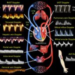
What can other venous Doppler waveforms tell us when the conventional #VExUS veins are inaccessible or difficult to interpret?
#POCUS #Nephpearls #FOAMed

Accessory mitral valve tissue!
#echofirst #POCUS #FOAMed
🔗Rabischoffsky A, et al. CASE, 2017; 2, 9-11