

professor at KAIST | cofounder/CTO of @TomocubeInc. | advisor, The Wave Talk Inc. | biomedical optics | microscopy | physics | waves | biotech
Daejeon, Republic of Korea
Joined July 2010
- Tweets742
- Following1.2K
- Followers783
- Likes1.4K



Join us in Hong Kong for the 6th QPI Symposium! 🇭🇰
📅 20 Nov 2026
📍 CUHK
🎤 Invited speakers: Nicholas Durr, Yuan Luo, Kevin Tsia, Kenneth K. Y. Wong Yongsoo Yang
📝 Poster abstracts due 30 Oct
🎟️ Free registration
Register: forms.gle/XYUY5wwuBPstPwc39
Info: bmol.kaist.ac.kr/qpi-symposi…
🤖 Made with AI

Deep inside a living heart organoid. Unstained. In 3D.
At @Tomocubeinc, we keep pushing beyond our own imaging limits.
Imaging depth held you back? Get in touch to test your organoids before our next product launches.
tomocube.com/send-your-organ…
#Organoids #Tomocube

Embryo selection today rests on 2D morphology and the embryologist's eye. #Holotomography lets us measure refractive index — and therefore dry mass — in 3D, in living oocytes and embryos, without labels or fixation.
Congratulations to Dr. Chungha Lee, @BKaist!
Proud of the team, and grateful to our partners at CHA Bundang, Avenues, and @Tomocubeinc .
#Holotomography #IVF #ESHRE2026 #KAIST

KAIST researchers developed a non-invasive 3D imaging approach that tracks live oocytes and embryos over time.
Dr. Chungha Lee received the ESHRE 2026 Basic Science Award.
#KAIST #IVF #ESHRE2026

📢 Call for Papers — Quantitative Phase Imaging XIII @SPIEtweets Photonics West 2027 (San Francisco, 30 Jan–4 Feb).
🗓 Abstract deadline: 22 July 2026 (site stays open ~1 week more)
👉 spie.org/BO506
submit your latest QPI work & share widely!
#QPI #BiomedicalOptics
Grateful to Joonseok Choi for featuring my work in Pressian’s “Frontiers of Science.” We discussed holotomography—3D, label-free imaging of living cells—and the journey from KAIST research to Tomocube.
pressian.com/pages/articles/…
#Microscopy #Biotech #Tomocube
Great to welcome @FredSangyeon back to my lab. From undergraduate researcher in my group to HMS faculty, he is now moving to Rice to expand pioneering work on ultra-small lasers for bio/medical applications. PhD candidates and early postdocs—reach out!
🤖 Made with AI

A heartfelt surprise at #OpticaBiophotonics26.
The Michael Feld Biophotonics Award ceremony was originally planned for September, but since I could not attend, @OpticaWorldwide kindly arranged a special moment during the plenary. Deeply grateful to optica and the committee.


Thrilled to announce our new publication! We introduce iDTT, a technique to map 3D optical “fingerprints” of materials. More stable than laser-based methods, it enables non-destructive 3D analysis for applications from materials science to biomedicine. nature.com/articles/s41566-0…

Congrats to the Mayo Clinic team on their Nature Immunology study showing SIRT2-mediated deacetylation of LCK (K228) sets TCR signaling magnitude.
nature.com/articles/s41590-0…

A compelling step toward quantitative immune signaling. Great to see @Tomocubeinc holotomography used for live-cell analysis. 👏
Honored to receive the 2026 Michael S. Feld Biophotonics Award from @OpticaWorldwide
Grateful to my students, collaborators, and colleagues at KAIST and Tomocube. Biophotonics continues to reveal new dimensions of life through light—especially in quantitative phase imaging.

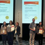
Great to see the next generation rising in Quantitative Phase Imaging at SPIE Photonics West (San Francisco).
🏆 Gabriel Popescu Award: Hyobin Cho (UT Austin)
🥇 Best Presentation Awards: Joshua F. Lerner (Duke) and Zih-Ying Fu (Georgia Tech)

Outstanding science, fair selection, and a powerful reminder of Gabriel Popescu’s legacy in empowering young researchers.
Please support the Gabriel Popescu Foundation 👉 gabrielpopescufoundation.org
#QPI #PhotonicsWest #YoungResearchers
New Cell paper from Markus Basan lab (Harvard Medical School) using @Tomocubeinc holotomography
Shows how label-free biomass density measurement unlocks quantitative biology in live cells.
Beyond imaging. once unimaginable is now experimentally possible.
#Tomocube

See the full text: cell.com/cell/abstract/S0092…
For more information or a consultation, please contact: [email protected]

Reviewed 64 HT papers published in 2025 and mapped where the field is heading.
HT has moved beyond niche imaging → now a label-free 3D phenotyping platform across cell biology, organoids, pathology & infection, increasingly powered by AI.
👉 arxiv.org/abs/2601.02611
