

Official Twitter account of the Palaeontological Association: https://xitter.cf/t.co/wUu2EEI1Tc. Re-posts of jobs and other events do not mean endorsement
- Tweets19.2K
- Following2.8K
- Followers12.5K
- Likes15.7K




ALT Digital reconstruction of the elements of the conodont Pseudofurnishius murcianus apparatus. At the top, the 15 elements are laid out; similar elements are shown in the same colour. A rectangular legend in the lower left of this area shows the alpha numeric code for each type of element. At bottom left is a model interpretation of how the elements were associated in life, in dextrolateral view. The dark blue rectangle at lower right is a cover image from the journal Palaeontology.


ALT In 1910, geologist Mignon Talbot (1869–1950) spotted bones in a sandstone block near Mount Holyoke College. In 1911 she named them Podokesaurus holyokensis, the first non-avian dinosaur named by a woman. The fossil burned in 1917.

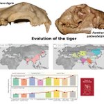
ALT Top, 2 photographs of tiger skulls; left, a modern Panthera tigris skull (AMNH M113743); right, a fossil, new subspecies Panthera tigris palaeobeijingensis (Zd00215) from Zhoukoudian, Beijing, earliest Middle Pleistocene. Photographs not to scale. Two grey boxes below contain maps showing the mainland distribution of the tiger lineage at two points in geological time: left, 0.4 to 0.013 million years ago, the pink area shows the distributon of Panther tigris; right, 0.013 million years ago to the present day, showing a number of different subspecies in different colours. Bottom: 2 histograms tracing the evolution of 2 different dental traits (changing morphology of 2 teeth). From left to right, the columns illustrate the condition in: in typical Panther palaeosinensis (from northern and southern China); transitional forms (2 columns); four Panther tigris subspecies. Red rectangle in lower right corner is a cover image from the journal Papers in Palaeontology.

ALT Images of a pair of right manus-pes (hand-foot) prints. The track form is named Pachychirotherium gardenaensis (UR 75/2, holotype). The top two images are photographs taken with oblique lighting; from the upper side on the left and from the right side of the right image. Bottom left is a multicoloured false-colour depth map of the same prints: dark red colours indicate a depth of 2 cm, dark blue zero depth. At the bottom right is a line drawing interpreting the outlines of the two prints; it has a 5 cm scale bar on the right. In the far bottom right corner of the image is a blue rectangle, which is a cover image for the journal Palaeontology.

ALT Photographs of rove beetles preserved in amber. Top left: Lathrobium rovnensis (holotype: IZAN K-8422), whole beetle, dorsal view with head to the top. Bottom left: Lathrobium athanasii sp. nov. (holotype: SDEI 217-3) in dorsal view, with a close-up view of the final sternite as reconstructed from micro-CT imaging (inset greyscale image). Top right: Nazeris nikitasi sp. nov. (holotype: NHDM Danish-amber-sp1): whole specimen in lateral view, with close up of the head and anterior body from micro-CT scans (inset greyscale image). Bottom right: Lathrobium arcanum sp. nov. (holotype: SDEI 719-3), head and anterior body shown in dorsal view. All images have a scale bar at the bottom, all scale bars are 0.5 mm. Small red rectangle (upper centre) is a cover image from the journal Papers in Palaeontology.

ALT Illustration of characters from the atlas vertebra distributed across the modern lissamphibian phylogenetic tree. Legend of characters and their respective symbols at top left. Underneath is a CT rendering of the atlas of Changchunerpeton nonganensis (RCPS VJ4001) in two views (whole specimen below, sagittal section above) and a black arrow indicating the placement of this taxon on the Hynobiidae lineage within the modern lissamphibian tree. Grey vertical bars on the tree highlight the Triassic, Cretaceous and Neogene time intervals (left to right). On the right, two columns of pairs of CT renderings of the atlases of different extant lissamphibians. In each case, the entire atlas in anterior view is shown on the left, and a rotated sagittal section showing the medial wall of the neural arch and the interior of the centrum in right lateral and slightly anterior view, on the right. Pink and blue arrows indicate the positions of the spinal cord and notochordal canal on the specimens.


ALT Upper left: Photograph of an articulated branch system of Lepidodentron aculeatum from the Bashkirian (Pennsylvanian, upper Carboniferous) of Hagen-Vorhalle, Germany (WMNM P85752). The specimen is approximately 350 cm long and about 200 cm wide (scale bar on the right is 100 cm). Lower left: Enlargement of part of the branch system: second ramification of the right main branch. Scale bar in lower right corner of photograph is 1 cm. On the right: an artist's reconstruction of one interpretation of Lepidodentron aculeatum (illustration copyright Stefan Sølberg; Mørkøv, Denmark, 2026). Red rectangle at bottom centre is a cover image from the journal Papers in Palaeontology.

ALT Circular schematic diagram summarising anatomical, dendrochronological and taphonomic evidence from the Lowenstein Formation (Norian, Bavaria). AT the top in blue, tree height is estimated at around 43 m. Next clockwise, in red, growth rings suggest alternating wet and dry seasons, but also episodic drought recorded by false rings. In green on the left, evidence of fungal decay (white rot and brown rot) suggest post-mortem saproxylic activity, causing decay of wood and nutrient recycling. All of this evidence suggests long-lived trees (over 500 years old) living in a dynamic climate-sensitive continental forest. Red rectangle in lower right corner is a cover image from the journal Papers in Palaeontology.


ALT Life reconstruction of Thoracosaurus isorhynchus hunting fish in a shallow marine environment. Underwater scene with the narrow snout of a crocodile-like animal, with a long tail, rising up from the sea bed to capture a small fish from a passing shoal in the centre of the image. Fish and shellfish in the foreground on the right. Artwork by Nathan Dehaut.


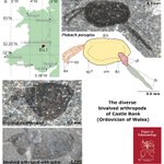
ALT Top left: map of Wales and the border with western England (in green); the location of the Builth-Llandrindod Inlier is shown in red (B-LI). Top right: photograph of the holotype and only known specimen of new species Ffabach peryglus from Castle Bank (Ordovician; NMW.2021.3G.12). Underneath is an annotated line drawing of the specimen with the carapace in yellow, the anterior appendages in orange to the left, and the posterior trunk (pink) and tail spine (green) on the right. Scale bars at the top right of the photograph and bottom left of the drawing, are both 0.5 mm. Lower left: two photographs of specimens currently without names: at the top is a new bivalved euarthropod (NMW.2021.3G.15i; scale bar at lower right is 0.2 mm); at the bottom is a bivalved arthropod carapace with spine (NMW.2021.3G.170; scale bar at lower right is 1 mm). Red rectangle in the bottom right corner is a cover image from the journal Papers in Palaeontology.


ALT Upper left: photograph of fossil specimen IVPP V26687, the placoderm Yipetalichthys maxiongensis, a natural mould of the dorsal part of the endocranium, preserving only a portion of the preorbital plate. Scale bar at lower right of photograph is 10 mm. Lower right, interpretive line drawing. Red rectangle in lower left corner is a cover image from the journal Papers in Palaeontology.

ALT On the left: aerial photo of Section 2 of the Duntulm tracksite, with a line drawing below, highlighting Trackway 1 (line below, closer to vertical) and Trackway 2 (line above, closer to horizontal). Scale bar between the two images is 2 m. Lower right: aerial photo and line drawing of Trackway 1 (scale bar at lower right is 1 m). Upper right: photograph and line drawing of a manus-pes pair of footprints, illustrating the difference in size observed in all tracks. The manus at the top (DUNT_26) is smaller than the pes (DUNT_27). Scale bar to lower right of drawing is 5 cm. Red rectangle at the bottom is a cover image from the journal Papers in Palaeontology.


ALT Plot showing the height in metres (vertical axis) against trunk diameter (horizontal axis) for nine populations of Pinaceae trees from Denmark, China, Turkiye and Mexico. Coloured lines shown the ten different models that were fitted to this dataset.

ALT Upper left: photograph of a yellow-brown rock sample containing a fragment of the postcranial skeleton of a reptiliomorph (?Nyctiboetus kassini) (darker brown) with cololite from the Shikhovo–Chirki locality (PIN 161/180). Scale underneath is 10 mm. Lower left: interpretative sketch of the specimen showing tetrapod remains in brown, fields of scales (paler brown), the cololite in grey and undigested remains in red. Upper right: tentative reconstruction of the fragmentary skeleton in dorsal view; the bones in red are preserved and the cololite is shown in blue. Lower right: reconstruction of the postcranial skeleton with cololite, based on CT data. Scale bar in lower right corner is 5 mm. Red rectangle on right side is a cover image from the journal Papers in Palaeontology.