

Manufacturer of scientific instrumentation for life science research & clinical use. And manufacturer of veterinary medical devices.
Joined February 2017
- Tweets1.3K
- Following95
- Followers301
- Likes110





Key Factors in Weak-Signal Detection for Small-Animal In Vivo Imaging
Same −90°C cooling. Same weak-signal detection performance?
Not necessarily.
In small-animal in vivo imaging, deep cooling helps reduce dark current. But cooling temperature alone doesn’t tell the whole story.
When evaluating a system, look beyond temperature:
• Quantum efficiency (QE): How efficiently does the detector convert photons into signal at your target wavelengths?
• Dark current: How much background accumulates under your actual operating conditions and exposure times?
• Read noise: Can faint signals be reliably distinguished from noise?
• Pixel binning: Does improved detectability preserve the spatial detail your study requires?
The most useful comparison starts with your experiment: test under matched conditions and assess weak-signal detection, quantitative reliability, and repeatability.
A brighter-looking image is only part of the picture. What matters is whether the signal can be reliably detected and measured.
#InVivoImaging #Bioluminescence #PreclinicalResearch #SmallAnimalImaging #ScientificImaging


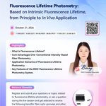
1/ RWD Lab Academy Webinar Series: Brand-New Content Now Live! Fluorescence Lifetime Photometry: Based on Intrinsic Fluorescence Lifetime, from Principle to 𝑰𝒏 𝑽𝒊𝒗𝒐 Application.
2/ Struggling with FLiP experiments? Probe expression variability, excitation power fluctuations, photobleaching, cross-brain-region/animal comparison, and absolute quantification?
3/ Join this RWD webinar to explore the Fluorescence Lifetime Photometry System—basic concepts, core advantages, typical applications, and reliable quantitative support.
4/ 📅 Oct 21, 2026 | 11:30 EDT / 10:30 CDT / 09:30 MDT / 08:30 PDT / 17:30 (CET) / 23:30 BJT
👩🔬 Speaker: Berry, RWD Solution Specialist
5/ Highlights: What is fluorescence lifetime? Core advantages over intensity-based fiber photometry? Application scenarios? Key features of the RWD FLiP system?
6/ Beyond skills: FLiP uses TCSPC for real-time monitoring in freely moving animals—high anti-interference, long-term stability, cross-sample comparison, absolute quantification.
7/ 🎁 Live interaction rewards.
Register via link/QR on poster:
us02web.zoom.us/webinar/regi…
#RWD #Webinar #FluorescenceLifetimePhotometry #Neuroscience #BrainResearch

Tracking Nanovaccine Delivery to Lymph Nodes with In Vivo Imaging
Where a nanovaccine accumulates—and how long it remains there—are key considerations in evaluating its delivery. In cancer nanovaccine research, tracking these changes over time helps researchers understand in vivo behavior and assess vaccine design.
The RWD MOIS HT Preclinical In Vivo Optical Imaging System has recently been installed at Hainan Medical University, supporting research into cancer nanovaccine delivery.
The research team is using the MOIS HT to track nanovaccine accumulation and retention in draining lymph nodes. Non-invasive fluorescence imaging across multiple time points allows researchers to follow signal changes in the same animal, providing a longitudinal view that conventional tissue sampling cannot readily offer.
Combined with region-of-interest (ROI) analysis, these images enable researchers to compare fluorescence signal intensity over time and visualize changes in nanovaccine distribution. The system’s imaging sensitivity and stable performance help maintain a high signal-to-noise ratio even under weak-signal conditions, allowing dynamic fluorescence changes to be captured clearly.
By making the delivery process visible and measurable over time, the MOIS HT provides imaging evidence to support the evaluation of nanovaccine delivery efficiency in vivo.
👉 Learn more about the RWD MOIS HT: rwdstco.com/product-item/sma…
#InVivoImaging #Nanovaccines #CancerResearch #DrugDelivery #Biodistribution #PreclinicalResearch #RWDLifeScience


New Launch: RWD M1416 High-Speed Benchtop Centrifuge (Ambient Model)
We're excited to announce that the RWD M1416 High-Speed Benchtop Centrifuge is now available in an ambient model for overseas markets.
🔹 Max speed 18,000 rpm (31,152 × g) | Capacity up to 4 × 400 mL
🔹 Flexible rotor options for 0.2 mL–400 mL samples
🔹 Self-locking rotors & quick-lock aerosol-tight lids
🔹 Rotor ID recognition, scheduling, touchscreen control
Fast, safe, and flexible — built for the modern lab.
DM us to learn more.
#RWD #Centrifuge #LabEquipment #LifeScience #NewProductLaunch

✨ That’s a wrap on #KSBNS2026!
Honored to have joined KSBNS 2026 as a Gold Sponsor and connect with the neuroscience community in Daejeon. 🧠
Thank you to everyone who visited RWD at Booth No.25-26, shared your insights, and connected with our team.
And thank you to the KSBNS team for bringing the neuroscience community together!
Until next time! 👋
@KsbnsNeuro
#Neuroscience #BrainResearch #RWDLifeScience


We're excited to be at #WMIC2026 and connect with researchers and experts from across the molecular imaging community!
Stop by Booth #303 to explore the RWD MOIS HT Preclinical In Vivo Optical Imaging System and exchange ideas on in vivo imaging applications.
We look forward to seeing you at Booth #303!
#WMIC2026 #WMIS #MolecularImaging #InVivoImaging #OpticalImaging #PreclinicalResearch


One Rotor, Infinite Possibilities – The New Multifunctional Rotor is now available globally!
We are excited to announce the global launch of the new multifunctional rotor!🌍
One rotor, adapted for virtually all your routine centrifugation needs.
Why it matters:
💰 Cuts costs – one rotor replaces two
⚡ Saves time – quick adapter changes
🛡️ Ensures safety – airtight lid prevents aerosol leakage
📦 Order now. Contact your local RWD representative to learn more.
#RWD #Centrifuge #LabEfficiency #MultifunctionalRotor #LifeSciences #NewProduct

From Tumor Progression to Micrometastases: Capturing Disease Dynamics with In Vivo Imaging
In cancer and disease research, biological processes such as inflammation, gene expression, and tumor metastasis evolve dynamically over time. Capturing these changes longitudinally in living animals can provide valuable insights into disease progression and help researchers evaluate therapeutic interventions.
Recently, the RWD MOIS HT Preclinical In Vivo Optical Imaging System was deployed at Sun Yat-sen University Cancer Center, providing in vivo imaging support for research teams investigating cancer and disease mechanisms.
In tumor metastasis research, early-stage micrometastatic lesions can produce weak signals that are easily masked by background interference, placing higher demands on imaging sensitivity and signal-to-noise performance. For the detection of subtle metastatic signals in the lungs, the RWD MOIS HT combines ultra-low background noise with efficient photon collection to capture and quantify weak bioluminescent signals, supporting the visualization of early micrometastatic lesions and the investigation of tumor metastasis.
👉 Learn more about the RWD MOIS HT: rwdstco.com/product-item/sma…
#InVivoImaging #CancerResearch #TumorMetastasis #Micrometastasis #PreclinicalResearch #RWDLifeScience


From Distribution to Dynamics: Advancing Exosome Research with In Vivo Imaging
Exosomes are increasingly being explored in drug delivery, disease research, and therapeutic development. Understanding their in vivo distribution, migration, tissue accumulation, and dynamic behavior is essential for advancing exosome research.
Shenzhen University of Technology is expanding its capabilities in this area with the deployment of the RWD MOIS HT Preclinical In Vivo Optical Imaging System, supporting ongoing research in exosome tracking, biodistribution, and delivery.
From Distribution to Dynamics:
Conventional endpoint-based analyses often require animals to be sacrificed at different time points for tissue collection, making it difficult to continuously monitor changes in the same animal.
In vivo imaging provides a non-invasive approach to longitudinally tracking labeled exosomes, enabling researchers to observe their dynamic behavior within living animals.
With fluorescence and bioluminescence imaging, the MOIS HT supports applications including:
• In vivo tracking – monitoring labeled exosomes across multiple time points.
• Biodistribution – visualizing changes in exosome signals across different tissues and regions.
• Tissue accumulation – assessing where and how exosomes accumulate over time.
• Delivery studies – providing dynamic data to evaluate in vivo delivery performance.
The MOIS HT combines a back-illuminated, high-quantum-efficiency CCD detector with high-sensitivity optical detection, supporting the capture and quantification of weak biological signals. Fluorescence signal separation further helps distinguish target signals from tissue autofluorescence, supporting clearer visualization and more reliable quantitative analysis.
👉 Learn more about the RWD MOIS HT: rwdstco.com/product-item/sma…
#ExosomeResearch #InVivoImaging #ExosomeTracking #Biodistribution #DrugDelivery #PreclinicalResearch #LifeScience #


🧠 #KSBNS2026 is underway!
Proud to be a Gold Sponsor and connect with the neuroscience community in Daejeon.
📅 September 6–8
📍 Booth No.25-26
📌 Daejeon Convention Center, Daejeon, Korea
Final day — come meet the RWD team!
@KsbnsNeuro
#Neuroscience #BrainResearch #RWDLifeScience


Another High-Impact Paper Featuring RWD Small Animal In Vivo Imaging System! 🎉
RWD small animal in vivo imaging system supported research teams from Guangzhou University of Chinese Medicine and Xidian University in publishing three high-impact papers in Biomaterials, Materials Today Bio, and Advanced Composites and Hybrid Materials. These studies cover intranasal brain-targeted delivery for Parkinson's disease, tumor metabolic reprogramming immunotherapy, and cuproptosis-enhanced radiotherapy.
While the three studies address distinct research questions, they all leverage the RWD MOIS HT system for critical applications—tracking drug delivery processes, monitoring nanomaterial biodistribution, and dynamically observing tumor-related signals—establishing a visual bridge between material design and therapeutic efficacy.
MOIS HT – The powerhouse behind high-impact research! 💪
#SmallAnimalImaging #InVivoImaging #Biomaterials #ParkinsonsResearch #CancerResearch #Nanomedicine #DrugDelivery #DrugTargeting #PreclinicalResearch #LifeSciences


🚀 4 Days to Go!
RWD is proud to be a Gold Sponsor of #KSBNS2026! 🧠
📅 September 6–8
📍 Booth No.25-26
📌 Daejeon Convention Center, Daejeon, Korea
Come explore our neuroscience research solutions and connect with our team!
@KsbnsNeuro
#Neuroscience #BrainResearch #RWDLifeScience

Struggling with background signals, signal attenuation, or probe selection in your in vivo imaging experiments?
In vivo optical imaging is a powerful tool for tracking physiological and pathological processes over time—yet experimental factors can significantly affect reproducibility and data quality.
To help biomedical researchers overcome these challenges, RWD has published the 📘 In Vivo Imaging Technology Whitepaper.
Inside the whitepaper:
🔹 Practical workflows: Model building ➔ Labeling ➔ Quantification ➔ Sources of error & troubleshooting
🔹 Real-world applications in Oncology, Drug Development, Exosome Tracking & Immunology
🔹 Ready-to-use appendix: Quick reference list for probes, substrates, and tracers
🔹 Guidance on imaging system selection & multimodal imaging strategies
Equip your lab with practical guidance to support more robust and reproducible imaging workflows.
📥 Get the full whitepaper now: [rwdstco.com/ebook/small-anim…]
#InVivo#InVivoImaging#MolecularImaging #Preclinical #OncologyResearch #DrugDevelopment #Exosome #Biotechnology #RWD##IVIS
Automated High-Viability Single-Cell Isolation from Mouse Brain: A Standardized Workflow
🧠dissect the tissue→ enzymatic digestion → filtering and centrifugation → debris removal → resuspension for later use—we’ve got every step covered for you.
All backed by these two "best partners" 🤝
⚙️ RWD Single Cell Suspension Dissociator –—30-minute rotary shearing during enzymatic digestion, stable temperature control, and efficient preparation of single-cell suspensions;
🧺 RWD High-Speed Benchtop Centrifuge —Stable, step-by-step sedimentation and washing, clear layer separation, and compact, non-loose cell pellets.
📌 Save this for your next run, and make the whole process smoother.
💡 Want the full specs and protocol kit? DM me for the complete data package 📦
#SingleCellSuspension #MouseBrain #TissueDissociation #CellIsolation #LabProtocol #Biotech #RWD #SingleCell #FlowCytometry #Neuroscience #LifeScienceResearch #CellBiology
Only 2 Days Left!
🧠 Live Webinar | Neurovascular Dysfunction in Brain Diseases:Mechanisms and Therapeutic Strategies - Insights from Ischemic Stroke and Alzheimer's Disease.
🗓️ August 27, 2026 ⏰ 7:00 PM KST/JST | 6:00 PM CST | 10:00 AM UTC
🎁 Bonus: Submit your questions for a chance to win MCAO sutures, surgical forceps, and precision scissors!
📱 Scan the QR code on the poster below to reserve your spot!
#Neuroscience #BrainResearch #IschemicStroke #AlzheimersDisease #LiveWebinar #RWDLifeScience

Struggling with common challenges in fiber photometry experiments — signal detection failure, loose ceramic ferrules, confusing 410 nm control channel principle, or unclear data readouts like ΔF/F & AUC?
RWD Lab Academy Webinar Session 2: Fiber Photometry: Step-by-Step Along the Experimental Workflow and Practical Tips
📅 September 2, 2026
🕒 11:30 EDT | 10:30 CDT | 09:30 MDT | 08:30 PDT | 23:30 BJT
👩🔬 Speaker: Ala – RWD Solution Specialist
✅ Full workflow from surgery to data analysis
🎁 Live exclusive: complimentary trial access + ceramic ferrules & surgical instrument giveaways
👉 Register via link or scan the QR code on the poster.
🔗us02web.zoom.us/webinar/regi…
#RWD #Webinar #FiberPhotometry #BrainResearch #Neuroscience #LifeScience #Neural Circuit

🧠 Two Brain Diseases. Two Neurovascular Mechanisms. One Webinar.
What drives neurovascular dysfunction in ischemic stroke and Alzheimer’s disease?
On August 27, join researchers from the Institute for Basic Science (IBS), Republic of Korea and Westlake University, China as they explore emerging mechanisms linking vascular dysfunction to brain disease.
🔬 From Ischemic StrokeDr. Jae-Hun Lee will investigate how reactive oxygen species drive astrocyte activation, fibrotic scar formation, and neuronal death, with a focus on the H₂O₂–FUT8–COL1 axis as a potential therapeutic target.
🧠 To Alzheimer’s DiseaseDr. Xing-Hua Quan will explore how impaired neurovascular coupling emerges as an early pathological feature of Alzheimer’s disease, and how cerebrovascular smooth muscle calcium oscillations may actively regulate neurovascular function.
📅 August 27, 2026 | 6:00 PM Beijing Time / 10:00 AM UTC🌐 Live Webinar | In English
📱 Scan the QR code on the poster to register and join the live webinar.
#Neuroscience #NeurovascularCoupling #IschemicStroke #AlzheimersDisease #BrainResearch #NeurovascularDysfunction #LiveWebinar #RWDLifeScience

📌 Live Webinar | Neurovascular Dysfunction in Brain Diseases:Mechanisms and Therapeutic Strategies - Insights from Ischemic Stroke and Alzheimer's Disease
🗓️ August 27, 2026 | 7:00 PM KST / 7:00 PM JST / 6:00 PM Beijing Time / 10:00 AM UTC🌐 Webinar Language: English
👨🔬 Special Guest: Prof. C. Justin Lee Director, Center for Memory and Glioscience, Institute for Basic Science (IBS), Republic of Korea
👨🔬 Speakers: Dr. Jae-Hun Lee | Senior Researcher, IBS Dr. Xing-Hua Quan | Doctoral Researcher, Westlake University
💡 Featured Reports:1️⃣ Ischemic Stroke — Exploring oxidative stress-induced astrocytic collagen biosynthesis and neuronal death, and identifying the H₂O₂–FUT8–COL1 axis as a potential therapeutic target.
2️⃣ Alzheimer’s Disease — Investigating early neurovascular coupling deficits and the regulatory role of cerebrovascular smooth muscle calcium oscillations.
📱 Scan the QR code on the poster to register and join the live webinar.
🎁 Submit your questions during registration or the webinar for a chance to win research tools, including MCAO sutures, precision scissors, and surgical forceps.
#Neuroscience #NeurovascularCoupling #IschemicStroke #AlzheimersDisease #BrainResearch #NeurovascularDysfunction #LiveWebinar #RWDLifeScience

We're excited to welcome you to RWD Booth 82-85 at NEURO2026! Explore our latest neuroscience research solutions, meet our experts, and discover how we're supporting laboratories worldwide. Visit us in Kobe—we look forward to connecting with you! #NEURO2026 #Neuroscience #Innovation #RWDLifeScience
Thank you for joining the RWD Neuroscience Networking Night!
It was a pleasure to bring together researchers and partners for an evening of scientific discussions, inspiring talks, and meaningful networking during NEURO2026.
Thank you to our speakers and everyone who joined us in Kobe.
#NEURO2026 #Neuroscience #RWDLifeScience


