

Since 1939, Hitachi has developed and manufactured various Electron Microscopes including unique tabletop and ultrahigh voltage as well as SEM, TEM and FIB.
Joined March 2018
- Tweets3.7K
- Following306
- Followers23K
- Likes1.9K



Hitachi Electron Microscope retweeted
It’s National Chemistry Week !
⚗️🧪⚛️🧬🥼🔬
(October 18-24, 2026)
TEM-EELS analysis of Gas atmosphere using FE-TEM with Gas Injection
Various gasses are now introduced for in-situ electron microscopy. EELS spectra of H2 gas, O2 gas and air were acquired near specimen and characteristic K-edge was clearly observed in each spectrum. Gas atmosphere can be precisely controlled for in-situ electron microscopy.
About Gas Injection, visit hitachi-hightech.com/global/…
Hitachi Electron Microscope retweeted
It’s International Infection Prevention Week ! 😷🚰🧴💉🧤
(October 19-25, 2026)
Gauze mask and Polyurethane mask
Masks are made to take advantage of materials' special properties and characteristics in order to block droplets and particles at the micrometer and nanometer level — whether that involves multiple layers of gauze with thick fibers, a non-woven material with an intricate mesh of extremely fine fibers, or polyurethane that catches particles using static electricity.
For more details, visit hitachi-hightech.com/global/…
It’s World Food Day ! 🥖🥚🍅🍌🥛🥩🐟
(October 16, 2025)
“Delicious foods are also structured beautifully. Exploring Food Structure to Establish the Science of Cooking”
Culinary science has a variety of approaches to research. Cooking methods, ingredient analysis, food properties, psychometrics (sensory evaluation), food culture, and food histology all comprise important parts of this research. Flavor and texture, such as hardness or chewiness, are all closely related to a food’s structure, and food histology has evolved along with techniques for observing tissue structure in order to examine the properties of each type of food.
For this interview, I sat down with Professor Machiko Mineki, who has been developing techniques for observing the microscopic structure of tissue, and has received many accolades. I asked Professor Mineki about progress in food histology research and what role electron microscopy plays in it.
Read the full article here: hitachi-hightech.com/global/…


It’s National Aesthetician Day ! 👐🖌🧴
(October 15)
Cryo SEM and Cryo TEM of Liposomes
Check out empty and drug-loaded liposomes here: youtube.com/watch?v=Tgc-m1g1…


It’s International E-Waste Day ! ♻📱🔋🔦🔌💡
(October 14)
IC Lead Frame inspection using large DOF (Depth of Focus) SEM
Uncoated IC chips on a printed circuit board (PCB: 12 cm x 10 cm) were observed at low accelerating voltage (1.0-2.0 kV). IC chips were three-dimensionally observed at 70 degree stage tilt. The height difference between the top and the bottom of the image was approximately 5 cm at very low magnification (x5). Micro cracks were recognized on the surface of Ni/Pd-plated lead frame.
About the large DOF (Depth of Focus) SEM, visit hitachi-hightech.com/global/…
Happy National M&M Day ! 🍫🍬
(October 13)
Cryo ion milling & low voltage SEM of chocolate
Chocolate under Electron Microscope !
Cross section of candy coated chocolate was prepared by cooled ion milling. Interface between chocolate and coating and the dispersion of sugar crystals and eggshell calcium were clearly observed without thermal damage.
About Ion Milling, visit hitachi-hightech.com/global/…
Happy World Algae Day ! 🏞️
(October 12)
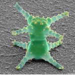
Micro Monster, a single-celled microalga, Staurastrum sp. prepared using Ionic Liquid
Although it may look like an industrial product, it is a single-celled microalga called Staurastrum sp.
It is characterized by its projections and a constriction in the center of the cell.
Staurastrum is found worldwide, but species with four projections on each end are rare.
By using an ionic liquid, we were able to photograph it without distorting its distinctive projections.
This work was presented at the "photo contest" hosted by the Japanese Society of Microscopy.
For more details, visit hitachi-hightech.com/jp/ja/k…

It’s National Metric Day ! 📏📐🔬⚖🌡🧪
(October 10)
Automated SEM imaging and measurement of resist line patterns
50 Resist line patterns were automatically searched, captured and measured using a combination of an SEM automation software, EM Flow Creator (EMFC), and an image processing software, Image Pro. After moving to the pattern position, EMFC image recognition function was used to fine-tune the position so that the line pattern is at the center of the image. SEM images were captured with the line pattern in the center. Image Pro’s macros were created to automates median filtering, binarization and line pattern width measurement. Automatic folder monitoring function enables the measurement as soon as images are acquired. The average value of the line width for the 50 patterns was 100.82 nm with three-sigma value of 3.68 nm.
About EM Flow Creator, visit hitachi-hightech.com/global/…
Happy National Nanotechnology Day ! 🔬🧬⚛️📱💻
(October 9)
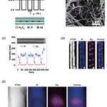
“Fabrication of nickel barcode nanowire and characterization by SU9000 scanning electron microscope”
In recent years, hollow metallic nanoparticles have attracted intense interest for their many remarkable properties, including giant specific surface area, catalytic activity, and low specific mass. Among their many applications are catalysts, sensors, energy-storage devices, molecular sieves, and batteries.1)To date, spherical nanoparticles,2) nanocubes,3)nanorods,4) and nanowires5) have been used as hollow metallic host nanoparticles. Nanowires are particularly promising host materials due to factors such as their strong adhesion to substrates and the ease with which they form network structures. Real-world applications of hollow nanowires include high-rate capacitors,6) solar cells,7)water purifiers,8) hydrogen producers,9) and catalysts.10)
One method of producing hollow nanowires is the template approach, which is capable of fabricating “barcode” nanowires with a periodic pore structure.11,12)In this method, electrochemical plating is carried out within a template in order to deposit a laminated structure containing two different metals: the metal that should be retained in the final structure (M1 in Fig. 1), and a sacrificial metal that is eventually dissolved to yield hollow regions (M2 in Fig. 1). Other reported fabrication techniques include those based on the Kirkendall effect13) and on galvano-substitution reactions.14)In all cases, precise control of the diameter and length of the host material, and of the hollow regions are important for obtaining the desired properties. In the present study, a structural characterization of such nanowires was performed by ultra-high resolution scanning electron microscopy (SEM) using a Hitachi High-Tech SU9000. Because the SU9000 allows—in addition to high-resolution SEM imaging—the acquisition of transmission images, dark-field images, and X-ray fluorescence (energy-dispersive X-ray spectroscopy) images within the same field of view, it is an extremely powerful instrument for investigating the exterior and interior of barcode nanowires, in additional to performing elemental analysis. In the experiments reported here, the full power of this instrument is demonstrated.
Read the full article here: hitachi-hightech.com/global/…


It’s National LED Light Day ! 💡🔦🚦🚨🚥
(October 7)
50 kV Micro-XRF of an LED chip on SEM
An LED chip was analyzed using 50 kV Micro-XRF on SEM. Fluorescent matters (Sr, Y) in encapsulating medium, LED (Ga) and silver electrode (Ag) at a depth of 700 μm were clearly visualized in the Micro-XRF map.
Available instrument: Hitachi Scanning Electron Microscopes SU3800/SU3900
Happy National Noodle Day !🍜🍝
(October 6)
SEM of Instant Noodles
Many instant noodles are processed by frying boiled noodles in oil.
The cavernous holes you see in the image are created by the evaporation of water when the noodles are fried in oil.
When you put the noodles in hot water, the water will fill these holes, returning the noodles to their original state so you can eat them.
Available instrument: Hitachi Tabletop Microscopes TM4000PlusIII/TM4000III


Hitachi Electron Microscope retweeted

Under an electron microscope,
the “same paper” can look completely different. 📄
When you look closer,
there are so many new discoveries to make. ˂( ╹ ‿ ╹ )˃💡
xitter.cf/Hitachi_ht_en/status/2…

The paper we use every day looks simple —
but under a microscope, its structure is completely different.
We compared these two:
Toilet paper
• Lots of gaps between fibres
• Breaks down easily in water
Paper wipes
• Thicker, more tightly tangled fibres
• Less likely to shed lint
Even though they are both paper,
their microscopic structures change depending on their purpose.
The closer we look at everyday things,
the more hidden design we discover.
If you found this interesting, let us know with a like!


It’s National Manufacturing Week ! 🔧🔨⚙⛑
(October 2–9, 2026)
#ElectronMicroscope #NationalManufacturingWeek #Drill
Cross-sectional analysis of Ultrahard Material (Cemented Carbide Drill)
7 mm wide cross section of ultrahard material, cemented carbide drill, was prepared in 5 hours by broad ion beam milling. While the whole shape was observed at low magnification, the fine laminate structure on the surface could be visualized at high magnification.
Available instrument: Hitachi Ion Milling System ArBlade 5000
Happy World Smile Day ! ☺😄😁☻ (^_^)/
(October 2, 2026)
“Smiling Pico-plankton”
This is a small organism of plankton collected in the northern Atlantic Ocean. The population of this plankton is high in cold water regions such as the Antarctic Ocean and the Arctic Ocean. People have very little knowledge about this plankton. Biological and ecological studies as well as other scientific investigation have just begun. Do you think that this plankton is smiling at you in the picture? It may appear in your dream tonight.
This work was presented at the "photo contest" hosted by the Japanese Society of Microscopy.
To know more about the image, check out the following post !

About the image, visit hitachi-hightech.com/global/…
Hitachi Electron Microscope retweeted

This scanning electron microscopy image shows extracellular web-like structures within a pulmonary embolic thrombus from a child with Mycoplasma pneumoniae pneumonia. doi.org/10.1172/jci.insight.…
Blood-clotting complications in pediatric pneumonia infection can be life threatening, but the underlying mechanisms of thrombosis have been unclear. Here, Huang et al. discover neutrophils promote thrombosis in #MycoplasmaPneumoniae pneumonia via neutrophil extracellular traps-mediated endothelial activation involving TLR2 and JAK–STAT3 signaling.


Hitachi Electron Microscope retweeted

They visited the following Departments / Laboratory of the Institute:
• Dept. of Pharmacology and Toxicology (With special reference to Transition Electron Microscope (TEM))
• Dept. of Pharmaceutics (With special reference to Scanning Electron Microscope (SEM))
and

