










ALT Sonographic greyscale imaging (a) along the anterior aspect of the right shoulder demonstrates the glenosphere (G) with a sinus tract (*) extending from the component toward the skin and communicating with a fluid collection (F) just deep to the skin (S). Color Doppler imaging (b) shows prominent hyperemia along the tract.


ALT History: 18-year-old male patient with intermittent hip pain for 5 years.


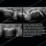
ALT Sonographic imaging along the dorsal aspect of the left hand reveals that the extensor tendons (dashed ovals) to the (a) middle, (b) ring, and (c) small fingers are subluxed ulnar at the level of the metacarpal head (M). (Right of image is patient's ulnar side.) Note the small finger has two extensor slops and the middle finger has trace tenosynovial fluid (*).


ALT History: 51 year-old-man with left sided low back pain. Surgery three months prior.

ALT Sonographic imaging of the left palm in the longitudinal plane in line with the fourth ray reveals a segmentally wavy course of the flexor digitorum profundus tendon to the ring finger (indicated by dashed line along the deep aspect of the tendon). Note the straight course of the overlying flexor digitorum superficialis tendon (dotted line is along the deep aspect of this tendon).


