
This is the official Twitter feed for Oregon Health & Science University's Casey Eye Institute.
Joined May 2012
- Tweets1.7K
- Following280
- Followers1.7K
- Likes1.1K


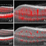




Does mitomycin C improve AGV outcomes? In this retrospective study, Casey Eye Institute researchers found that adjunct mitomycin C improves short-term but not long-term outcomes after Ahmed Glaucoma Valve surgery. mdpi.com/3443134 @Bioeng_MDPI

OHSU Casey Eye retweeted

Yali Jia @YaliJiaOCT et al. from Casey Eye Institute @CaseyEye quantify choriocapillaris flow deficits using projection-resolved optical coherence tomographic angiography and evaluate whether they are correlated with diabetic retinopathy.
doi.org/10.1167/iovs.66.12.1…

OHSU Casey Eye retweeted

2023 #LaskerLaureate David Huang tells us what keeps him going in this edition of #AskAScientist. @CaseyEye @OHSUNews
Researchers at Casey Eye Institute find novel #OCT-based optoretinography technology can evaluate larger retinal areas than previous methods & may be clinically useful in detecting and monitoring cone dysfunction in degenerative retinal diseases. iovs.arvojournals.org/articl…
Voting is open for Best of the Gorge 2025! We are honored receive a nomination from our community in the Health and Wellness category for providing excellent care to our patients in Hood River and The Dalles. Vote now through July 30! bestofthegorge.com

OHSU Casey Eye retweeted

Our collaborative work between @CaseyEye and @hkumed showing a potentially more accurate way for glaucoma detection and monitoring using individualised baseline retinal nerve fibre thickness and vascular patterns - Ophthalmology Science ophthalmologyscience.org/art…

Great expert panel discussion by @asrsdocs about the remarkable story of innovation and growth of optical coherence tomography #OCT with leading pioneers, including Casey Eye Institute's Dr. David Huang.

Just dropped: Dr. Christina Weng hosts retina legends Drs. Fujimoto, Huang, Duker & Sarraf for a panel discussion tracing the groundbreaking history of OCT — a technology that revolutionized the field. Watch now: youtu.be/Lf1ckPwVQnI?si=o_IY…. Explore more: HistoryofRetina.org
Join us June 21 to meet the experts behind some of the breakthroughs that are changing how we understand and treat eye disease. Learn about innovations in gene therapy, imaging, stem cells, AI & more.
Register: cvent.me/0kkWnx #ohsu @OHSUNews

OHSU Casey Eye retweeted

In the New Frontiers in Optical Coherence Tomography Issue bit.ly/3u6vw7v, David Huang et al. @CaseyEye study the Anatomy of the Macular Ganglion Cell Layer Plexus on Projection-Resolved Optical Coherence Tomographic Angiography, tvst.arvojournals.org/articl….

Join us for an inside look at Casey Eye Institute’s groundbreaking research! Timely Topics: Live from Marquam Hill — Casey Eye Institute; June 21 from 1-2:30 p.m.; OHSU Marquam Hill Campus, Old Library Auditorium; Register: cvent.me/0kkWnx
@OHSUNews

Researchers in Casey's #COOLLab refined their work on basic anatomical and functional characteristics of the ganglion cell layer plexus (GCLP) in the macula. Mapping of the GCLP may be useful in evaluating optic neuropathies. tvst.arvojournals.org/articl…

OHSU Casey Eye retweeted
Hiroshi Ishikawa et al. @CaseyEye @Wills_Eye study Normative Variability in Retinal Nerve Fiber Layer Thickness: Does It Matter Where the Peaks Are? tvst.arvojournals.org/articl…

Congrats to @OHSUNews's Dr. David Huang for induction into the @InventorsHOF Class of 2025! He co-invented optical coherence tomography #OCT, which has transformed eye care & improved the detection & management of eye diseases. invent.org/inductees/david-h… #NIHF2025 #ohsu

ALT A man stands in front of a large banner with his photo and name.
OHSU Casey Eye retweeted

Nice work on glaucoma:
Matrix stiffness regulates traction forces, cytoskeletal dynamics, and collagen reorganization in trabecular meshwork cells, potential for improved understanding of glaucoma pathogenesis.
@CaseyEye #NEIfunded
@CellPress paper: bit.ly/4jdVElq

ALT Illustrative diagram comparing a normal or glaucomatous human eye with a red-labeled type I collagen gel embedded with green fluorospheres simulating the eye's tissue stiffness. The diagram highlights parts of the eye such as the optic nerve head, ciliary muscle, conventional aqueous outflow pathway, and sclera. Insets show details of cellular responses in gels with varying stiffness.