

Advanced imaging research facility in the School of Biological Sciences at the University of Cambridge https://xitter.cf/t.co/8mP57CqBJW
Cambridge, England
Joined November 2018
- Tweets30
- Following114
- Followers430
- Likes13



A great opportunity in Cambridge to lead the Advanced Imaging Centre at the School of Biological Science ... 10 more days to apply
jobs.cam.ac.uk/job/40627/ @Cambridge_uni @cambridge_sbs @RoyalMicroSoc @EuroBioImaging @focalplane_jcs
Cambridge Advanced Imaging Centre retweeted

Check out our latest publication 'Visualising an invisible symbiosis' in @plantspplplanet! @plantsci @CropSciCentre @niabgroup
nph.onlinelibrary.wiley.com/…
October image of the month is from @Ruby_Peters_ of the
@PaluchLab and shows HeLa cells, labelled with Tubulin-GFP. Taken using our Elyra 7 Lattice SIM and colour coded for depth

Beautiful!

Excited to process and analyse my first nlsABACUS #lightsheet images, even if it is just the negative control😅! Martin Lenz at @CamMicroscopy and @slcuplants has built a brilliant microscope and is an imaging ninja.
Cambridge Advanced Imaging Centre retweeted

how to cover yourself when you are a plant? Just weave your own fabric! Check our collaborative work between @slcuplants @CUBotanicGarden @ChemCambridge @CamMicroscopy on how Dionysia tapetodes extrudes flavones threads from its glandular trichomes. biorxiv.org/content/10.1101/…

Cambridge Advanced Imaging Centre retweeted

Via #OSA_Optica: Single molecule light field microscopy ow.ly/Urg550BEkQ4 #FlatOptics #SpectralFIlters @koholleran @Cambridge_Uni @UniversityLeeds

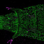
August image of the month: Drosophila larvae expressing green and magenta fluorescent proteins in nociceptive (noxious touch), and proprioceptive (body movement) neurons. Image: @Paul_brooks who researches neuron degeneration, captured by confocal microscope in @CamZoology

We're hiring! Research associate position in advanced photo-manipulation microscopy. Come join us in developing technology critical for cutting edge optical trapping, optogenetic and photoablation experiments. jobs.cam.ac.uk/job/26323/
Congratulations to @r_r_sims @sohaib_ar and the whole team for their new 3D super-resolution method, now published in Optica.
This Post is from an account that no longer exists. Learn more
July image of the month. Puromycin assay on cultured hippocampal neurons showing new proteins that are produced within 15 minutes (yellow/green) and microtuble-associated protein 2 staining (red).
Captured on our laser scanning confocal microscope by Dr Tanja Fuchsberger

The EPSRC Research Software Engineer Fellowship call is now live:
epsrc.ukri.org/funding/calls…
The @SarrisLab made incredible use of our two-photon microscope in their recently published research on neutrophil migration to sites of tissue damage cell.com/current-biology/ful…

In this video, we capture neutrophil swarming at a site of acute wounding, as the locus is invaded by opportunistic bacteria @CamMicroscopy
Hot off the press: our research lab's preprint of their new super-resolution microscopy technology.
This Post is from an account that no longer exists. Learn more
CAIC is starting a monthly series of informal clinics for super-resolution fluorescence microscopy. Come along for advice on getting the most out of our SIM, STED and SMLM systems. First clinic is Friday 21st February - follow link for details caic.bio.cam.ac.uk/news/supe…

Cambridge Advanced Imaging Centre retweeted

We got the cover! The wonderful @JessieJRH's new paper is out now in @FEBSJournal, with an excellent commentary from @WendyIngman. If you like immune cells and boobs, or nice 3D imaging, this is worth a read. febs.onlinelibrary.wiley.com… with @katevetpath & @cjwhelix

Fantastic opportunity to join the @EmilianiLab and get involved in advanced optogenetics technologies. You'll also get to work alongside the talented @r_r_sims who developed light sheet and light field microscopes in CAIC.

PhD Position in the #interdisciplinary PhD program #zenith_etn. @EmilianiLab is looking for candidates interested in non-linear optics,wavefront shaping and optogenetics( zenith-etn.com/projects/all-… ).Send your application soon,deadline is January 5th 2020! #ITN @MSCActions #zenith

Great work by Martin Lenz showcased in his talk about the multi-view light sheet microscope for gentle fast imaging of plants he has developed in the @slcuplants imaging facility.


Flaminia Kaluthantrige Don, a @BBSRC CASE student hosted by CAIC and Meri Huch's lab, used our two-photon microscope to great effect. She optimised imaging conditions in order to observe single cells proliferating into liver organoids over three days.
Congratulations to Hugo Poplimont who won a @RoyalMicroSoc prize for best use of microscopy in a presentation at #BSI2019. You can read about his work on neutrophil swarming in damaged tissue using our two-photon microscope in their recent @biorxivpreprint biorxiv.org/content/10.1101/…
