

The Archives of Pathology & Laboratory Medicine. An official publication of @Pathologists. Celebrating 100 years 🎉. Editor-in-Chief: Alain C. Borczuk, MD
- Tweets1.9K
- Following340
- Followers10.5K
- Likes898




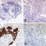


ALT Figure 1. Pleomorphic adenoma (A and B) and adenoid cystic carcinoma (C and D) showing high transcriptional repressor GATA binding 1 (TRPS1) positivity with stronger staining in abluminal cells. Cases of secretory carcinoma (E and F), acinic cell carcinoma (G and H), mucoepidermoid carcinoma (I and J), and hyalinizing clear cell carcinoma (K and L) showing diffuse strong TRPS1 staining Figure 3. Thyroid follicular carcinoma (A and B) showing high transcriptional repressor GATA binding 1 (TRPS1) staining (B); normal thyroid (C) also shows TRPS1 positivity; thyroid papillary carcinoma (D and E) showing high TRPS1 (E); clear cell renal cell carcinoma (F and G) with high-positive TRPS1 (G); TRPS1 staining in normal kidney (H); lung carcinoma (I through K) with intermediate-positive TRPS1 in papillary-acinar (J) and high-positive TRPS1 in signet ring (K) adenocarcinoma; colon (L and M) and periampullary (N and O) adenocarcinoma are negative for TRPS1


