

American Registry of Pathology Press: Home of the AFIP Fascicles
- Tweets1.7K
- Following1.1K
- Followers5.9K
- Likes286
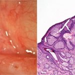




ALT SEBACEOUS GLAND HYPERPLASIA: CARUNCLE Left: A yellow, lobulated, well-circumscribed caruncular mass. (Courtesy of Drs. J. Shields and C. Shields, Philadelphia, PA.) Right: Hypertrophic and hyperplastic sebaceous gland lobules are arranged around a central dilated duct (arrowhead) (H&E stain).

ALT SMALL CELL CARCINOMA OF THE OVARY, HYPERCALCEMIC TYPE A: The tumor grows in a solid fashion with multiple pseudofollicles. B: The tumor cells are small with minimal cytoplasm. Numerous mitoses are identified. C,D: The pseudofollicles are lined by malignant cells, and are filled with an eosinophilic material. E: This image highlights the large cell variant of small cell hypercalcemic type, and demonstrates the rhabdoid like appearance of the tumor cells. F: Immunohistochemistry with BRG1 is diagnostic, with loss of this marker in the malignant cells (and retained staining in lymphocytes and other benign stromal cells).


ALT RETINOBLASTOMA: EXTENSIVELY NECROTIC A: CT image of a 2-year-old child who experienced eyelid edema and inflammation, accompanied by conjunctival edema (chemosis), pain, and proptosis. The cornea was opaque. The center of the proptotic eye contains bright white densities consistent with calcium. B: T2-weighted magnetic resonance imaging (MRI) demonstrates irregular densities in the vitreous cavity of the right eye in contrast with the even water-type opacity seen in the vitreous in the left eye. C: T1-weighted MRI illustrates the enhancement of the periocular tissues. D: Enucleated right eye after fixation in the same patient is remarkable for extensive tumor necrosis (TN), which is tan-yellow and soft alternating with bright white calcification in the anterior central vitreous cavity. E: The anterior portion of the eye shows the congested episcleral vessels, hazy cornea, and anterior chamber with dispersed pigment and blood.

ALT RETINOBLASTOMA A: Medium to large cells with high nuclear to cytoplasmic ratio, granular “salt and pepper” type chromatin, and scant delicate eosinophilic cytoplasm (retinoblasts). Mitotic figures and apoptotic cells are seen. B: Low-power view shows large geographic areas of necrosis and foci of dystrophic tumor cell calcification (Ca++). C: Viable tumor cells form a cuff around tumor vessels. The periphery of the cuff blends into areas of tumor necrosis. This typically occurs over 100 µm from the tumor vessel (see measuring bar). D: The basophilic deposits in vessels represent DNA fragments released from necrotic cells (Azzopardi phenomenon).

ALT DIFFERENTIATION IN RETINOBLASTOMA A: Flexner-Wintersteiner rosettes, typically seen in retinoblastoma, are composed of neoplastic cells with long cytoplasmic processes that converge upon a central lumen. The lumen appears to be bordered by basement membrane material, similar to the terminal bars of photoreceptors. B: Homer Wright rosettes have cytoplasmic processes filling the center of the structure and these rosettes may be seen in other neuroblastic tumors. C: Photoreceptor or neuronal differentiation is seen in very well-differentiated tumors (retinoma or retinocytoma). Mitoses and apoptosis are not seen in the well-differentiated fleurettes. Fleurettes are distinctive for well-formed photoreceptor-like structures with eosinophilic cytoplasm. D: Poorly differentiated retinoblastoma lacks any rosettes and mitotic figures are present (upper left corner).


ALT FIBRONECTIN GLOMERULOPATHY - H&E: Massive accumulation of amorphous, pale, eosinophilic material in the capillary walls and mesangium, obliterating most capillary lumens. This produces a lobular pattern of consolidation. The mesangial cell number appears increased, although distinct mesangial regions are no longer discernible.

ALT FIBRONECTIN GLOMERULOPATHY - PAS: The massive fibronectin deposits are weakly PAS-positive.

ALT FIBRONECTIN GLOMERULOPATHY- trichrome: These fibronectin deposits show focal fuchsinophilia (red staining) at their periphery with the Masson trichrome stains. Other examples may be more diffusely fuchsinophilic.

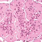
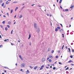
ALT LUPUS NEPHRITIS CLASS IV WITH LUPUS VASCULOPATHY: The lumen of this arteriole is occluded by eosinophilic material that consists of fibrin (darkly eosinophilic material internal to the swollen endothelium) and immune deposits (paler and more smudgy, homogeneous eosinophilic material between the fibrin and the media). There is no evidence of inflammatory infiltration of the vessel wall. Thus, this lesion is best termed a “vasculopathy,” and not a form of vasculitis. (H&E stain)

ALT LUPUS NEPHRITIS CLASS IV WITH LUPUS VASCULOPATHY: The intimal and intraluminal deposits in this small artery stain for IgG by immunofluorescence. Staining for fibrin/fibrinogen was also seen admixed with the immune deposits.

ALT IMMUNOGLOBULIN A (IgA) NEPHROPATHY: MESANGIAL DEPOSITS: Immunofluorescence microscopy on frozen tissue (A) and immunoenzyme microscopy on paraffin-embedded tissue (B) demonstrate intense mesangial staining for IgA.

ALT IgA NEPHROPATHY WITH MESANGIAL HYPERCELLULARITY: This glomerulus has expanded mesangial matrix and prominent global mesangial hypercellularity, exceeding three mesangial cells per mesangial area in multiple segments. The glomerular capillary lumina remain patent. (Hematoxylin and eosin [H&E] stain)

ALT IgA NEPHROPATHY WITH MESANGIAL HYPERCELLULARITY: This PAS-stained glomerulus has segmental foci of mesangial hypercellularity and matrix expansion (examples are seen at the 3 o’clock and 10 o’clock positions). These foci have greater than three mesangial cells in a single sector of matrix and are peripheral mesangial segments, not adjacent to the vascular stalk or hilus. If the majority of scorable glomeruli show changes similar to those pictured in figure 11-4, then a score of M1 would be awarded in the Oxford classification.


