

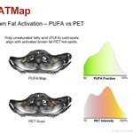





#BodyDigital #BATMap: Do you have active brown fat?🔥 In "browned” #fat, PET scan hotspots match #MRI poly-unsaturated fatty acid (PUFA) coldspots — suggesting a transition from PUFA enrichment to depletion as brown fat becomes fully active.

#BodyDigital #BATMap: "Browned" #fat — the metabolically active fat that burns energy 🔥 — isn't just one tissue. Fatty-acid composition mapping by #MRI reveals distinct brown and beige components within canonical brown fat depots — each with its own peak in total fat fraction.

Consistent #LiverMap results a week apart. Reliable imaging, actionable metrics. Thanks for sharing your healthy scan with #BodyDigital. 🎯

💃🏽My liver is healthy & happy… 😎I can see it! #LiverMap @bodymapdigital

ALT Scan - NUS August 2026 Singapore
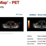
🔥 What is brown adipose tissue (#BAT)? It's active, fat-burning tissue that drives energy expenditure, linked to #Metabolism & healthy #Ageing.
⭐ #BATMap by #BodyDigital makes this hidden biology measurable using #MRI— no ionising radiation, ~10 minutes to track #interventions.

#BodyDigital #LiverMap demonstrates how #MRI multi‑component relaxometry can quantify #fibrosis, #inflammation, and #steatosis in fatty #liver disease from a ~10‑minute scan.
Terrific collaboration across Brazil 🇧🇷and Australia 🇦🇺.
sciencedirect.com/science/ar…

#BodyDigital #BATMap: In contrast to PET, BATMap isolates canonical “browned” #fat depots and maps brown→beige→white fat phenotypes — no tracer, no ionising radiation. Brown fat cells burn stored fats to generate heat, supporting glucose control and healthier metabolic balance.

#BodyDigital #Fat Metabolism: Brown fat has higher polyunsaturated fatty acids (PUFA) — a hallmark of healthier metabolic function. Yet in #MRI, this biochemical shift nudges the fat signal by only ~1%, hidden in the bis‑allylic fat spectral peak. #BodyMap is up to the challenge.

Body Digital retweeted

😍 Time to ‘See’ and ‘See the Time’ ....🥳 #ISMRM2026 linkedin.com/feed/update/urn…

🧠 #BodyDigital #BrainMap: Extra-Axonal Proton Density (EAPD) measured by #MRI has a relatively stable peak in healthy adults, with peak broadening emerging as a hallmark of #aging. 'Lifetime-weighting' on the peak resolves EAPD into a rising predictor of biological #brain age.⌛️

ALT Log-linear relationship fitted to pilot data on 17 cognitively normal volunteers and 5 Alzheimer's patients for "lifetime-weighted" extra-axonal proton density versus age (fit R-squared is 0.97, with some data outside age range shown).
These #BodyDigital #BrainMap reports, left to right, show insula matures later, with #axonal proton density (PD) giving stronger age-descrimnation than #myelin sheath PD. Younger brains also show a lower, tighter R2* peak, reflecting less iron load.🧠Now, who can guess the ages?


#BodyDigital #BrainMap: When metrics match and distributions align, it's the pattern that speaks. Subtle #brain #aging effects - like sulcal widening in the insula - emerge in the micro-architectural maps as changes in extra-cellular space, myelin density, and axonal integrity.🧠

ALT 47 years old


#BodyDigital #BrainMap: making neurodegeneration visible, in color. Healthy aging brains look "filled-in" —bright axons, intact #myelin, muted #inflammation. In #Alzheimers disease those colors fragment — clear regions expand as visible gaps in tissue volume. #MRI #BrainHealth 🧠


#BodyDigital #BrainMap: early onset #Alzheimer's versus an age-matched healthy volunteer. Neuro-degeneration presents with elevated #inflammation, reduced #myelin content and loss of #axon integrity in frontal lobe white matter, with increased #plaques evident in the putamen.


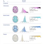
#BodyDigital #BrainMap: Mapping #infalmmation, #myelin content, #axon density, and #plaques in under 10 minutes scan time. Here, a baseline BrainMap report for a healthy volunteer, showing ROIs for frontal lobe white matter myelin, and an absence of plaques in the putamen (+). 🧠

#BodyDigital #LiverMapPlus: Intracellular #T2 mapping reveals #cancer #cell activity. #Liver #metastases show elevated T2 from healthy, increased T2 heterogeneity, and clear necrotic regions—reflecting increased membrane permeability and mobile water in #malignant cancer cells 🔬


#BodyDigital #LiverMap: In a setting of very high #liver #fat, before sequestration capacity and hepatocyte function decline, preserved cells can still mount a strong #inflammation response alongside advancing #fibrosis — a profile of active, progressive #NASH 🧐

#BodyDigital #LiverMap: With persistently high levels of #liver #fat, progressive fibrosis can alter the liver’s capacity to mount a robust inflammatory ‘healing’ response, so that only mild #inflammation may be observed in the setting of advance #fibrosis. 🔎

#BodyDigital #LiverMap: Regional increase in #liver #fat in fatty liver disease is associated with increased fibroinflammatory activity above normal (here in the right lobe of the liver), even when global measurement of #inflammation and #fibrosis is within normal.🧐

#BodyDigital #LiverMap: Sensitivity and reproducibility are essential to understand when our #liver is healthy and when it is not, no matter where we have an #MRI.

