

We build sophisticated #cell-electronic interfaces to probe intelligent biological networks by linking them to software. #Drugdiscovery
Pfäffikon SZ, Switzerland
Joined February 2015
- Tweets847
- Following514
- Followers858
- Likes528







What if dying neurons help build the retina?
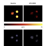
In a new publication from Savage and colleagues, the team uncovers a fascinating link between spontaneous neural activity, cell death, immune cells, and blood vessel growth in the developing retina.
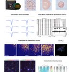
With the help of all 4,096 simultaneous recording electrodes, and the large recording area of CorePlate™ 1W 51/81, they were able to record from the activity of the entire retina. Combining this with large-scale calcium imaging, single-cell RNA sequencing and immunohistochemistry, the team mapped spontaneous retinal waves across the entire retina.
They discovered that wave initiation follows a structured centrifugal expansion, challenging long-standing assumptions that these early neural waves occur randomly. They showed that dying retinal ganglion cells are not passive, and that they actively direct development, generating local retinal waves and creating local hypoxia that guides blood vessel growth, signal microglia to engulf the dying cells and form distinct autofluorescent cluster complexes.
Together, the study presents a new framework in which neural activity, programmed cell death, immune responses, and vascular development are coordinated, ensuring that neural circuits and their supporting blood supply mature in synchrony.
Congratulations to Michael A Savage, Cori Bertram, Jean de Montigny, Courtney A Thorne, Rachel Queen, Majlinda Lako, Gerrit Hilgen, Evelyne Sernagor on this publication in eLife.
Read the full paper here: elifesciences.org/reviewed-p…
#3Brain #HDMEA #Electrophysiology #Retina #Neuroscience #Microglia #Angiogenesis #DevelopmentalBiology #NeuralDevelopment #VisionScience

Building a neuronal bridge across a broken spinal cord.
In a new publication from Wei et al., the team presents a novel approach for developing human induced pluripotent stem cell (hiPSC)-derived neuronal tissueoids (Ntoids) as improved cell-based treatments for spinal cord injury (SCI). The study demonstrates how functional neuronal tissueoids can serve as a neuronal relay, helping to reconstruct damaged neural pathways and restore motor function following complete spinal cord injury in rats.
To characterize the functional activity and network connectivity of the engineered neuronal tissueoids, the team used the HyperCAM Alpha paired with the CorePlate™ 6W-3D, which features 13,824 3D simultaneously recording electrodes across all six wells. Their findings demonstrated robust neuronal activity and functional network formation, confirming the tissueoids' maturation and functionality prior to transplantation.
Following transplantation, the neuronal tissueoids showed remarkable donor cell survival, innervation of the injury site, and partial integration with host neural circuits, with treated animals exhibiting improved motor recovery and electrophysiological performance. Further to this, in a separate experiment, the authors also demonstrated the ability of the neuronal tissueoids to form synaptic connections with human brain organoids.
Congratulations to Qing-shuai Wei, Quan-ying Xing, Yuan-huan Ma, Ke-jun Zhang, Wen-tao Ma, Mei-ling Zhang, Hui-juan Shi, Li-zhi Peng, Jun-hua Wang, Qi-xing Hu, Hou-ge Li, Ying Liu, Bi-qin Lai, Ge Li, Ying Ding, Yuan-shan Zeng, and Xiang Zeng on their publication in the Chemical Engineering Journal.
Read the full paper here:
doi.org/10.1016/j.cej.2026.1…
#3Brain #Neuroscience #CorePlate #HDMEA #StemCells #iPSC #Organoids #SpinalCordInjury #Electrophysiology #RegenerativeMedicine

Interested in hiPSC disease modelling? Our new Application Note is now available: High-Resolution Electrophysiological Profiling of Healthy and Dravet Syndrome Human iPSC-derived Neural Spheroids on CorePlate™ HD-MEAs.
Human iPSC-derived neural spheroids are powerful 3D models for studying neuronal function, disease mechanisms, and neuropharmacological responses. In this application note, we show how CorePlate™ enabled HD-MEAs enable high-resolution functional characterization of healthy and SCN1A heterozygous knockout Dravet syndrome neural spheroids from @FUJIFILM Cellular Dynamics.
By evaluating spontaneous and pharmacologically modulated activity, we reveal genotype-dependent differences in baseline activity, bursting behavior, network characteristics and functional connectivity, demonstrating the power of HD-MEAs for disease modeling and drug discovery.
Whether you're investigating neuronal function, exploring disease models, or screening therapeutic candidates, this application note highlights how CorePlate™ enabled HD-MEAs provide a powerful platform for functional assessments of hiPSC 3D neural models.
Check out the application note: resources.3brain.com/f1/down…

Final Call, Applications Close Today!
Don’t miss your chance to apply for the 3Brain - Massimo Grattarola Award:
• €15,000 to support your neuroscience research
• Free to apply
•Quick & easy application process
• Deadline: 17:00 CEST today, October 1, 2026
Apply now: 3brain.com/the-3brain-massim…
🤖 Made with AI

Applications for the 3Brain Massimo Grattarola Award close tomorrow!
Don't miss your chance to apply for the €15,000 award.
It's easy, free, and quick to apply.
Apply now: 3brain.com/the-3brain-massim…
#Neuroscience #Organoids #Spheroids #ResearchFunding #3DCellCulture #ResearchSupport #3Brain #HDMEA

Only 3 days left to Apply for the €15,000 Award!
Applications for the 3Brain – Massimo Grattarola Award close on October 1, 2026.
Its easy, free and quick to apply for! 3brain.com/the-3brain-massim…
#Neuroscience #Organoids #Spheroids #ResearchFunding #3DCellCulture #ResearchSupport #3Brain #HDMEA

Last Chance to Apply for the €15,000 Award!
Applications for the 3Brain – Massimo Grattarola Award close on October 1, 2026.
Win €15,000 towards your neuroscience research!
Its easy, free and quick to apply for!
👉3brain.com/the-3brain-massim…
#Neuroscience #Organoids #Spheroids #ResearchFunding #3DCellCulture #ResearchSupport #3Brain #HDMEA

The 2026 SPS Annual Meeting is here! We're excited to discuss the latest advances in neuronal and cardiac functional screening and how this is helping to drive safer drug development. Join us and the global safety pharmacology community in Montréal, Canada.
As novel therapeutic modalities and increasingly complex in vitro models continue to transform drug development, high-quality functional data is more important than ever. Whether you're evaluating compound safety on iPSC-derived neuronal and cardiac cells or investigating responses in advanced 3D models such as brain organoids, 3Brain's CorePlate™ enabled HD-MEAs deliver high-resolution, label-free functional insights in formats up to 96 well, helping researchers generate robust and scalable data for preclinical safety assessment.
Stop by Booth #2 to learn how HD-MEA technology is enabling more predictive and informative safety pharmacology workflows.
See you in Montréal!
#3Brain #HDMEA #SPS2026 #SafetyPharmacology #DrugDevelopment #PreclinicalResearch #Neuroscience #Cardiotoxicity #Organoids #DrugSafety #HighThroughputScreening

Measuring Function, Revealing Dysfunction: HD-MEAs in ALS Research
Understanding the cellular and network-level mechanisms underlying neurodegenerative diseases such as Amyotrophic Lateral Sclerosis (ALS) is fundamental to advancing our knowledge of disease progression and neuronal dysfunction.
High-Density Microelectrode Array (HD-MEA) technology such as that found in CorePlate™ is providing researchers with high-resolution insights into neuronal activity and network dynamics, helping to reveal disease-relevant phenotypes and evaluate potential therapeutic approaches.
This article highlights how HD-MEAs can support neurodegenerative disease research, including ALS, by providing powerful functional readouts that complement molecular and cellular analyses.
If you're interested in neurodegenerative disease research, drug discovery, or advanced electrophysiology, this article from Zehra Yagmur Erol, Christopher Quintanilla and P. Hande Ozdinler is well worth a read.
Check out the article here: febs.onlinelibrary.wiley.com…
What do you see as the biggest opportunities for HD-MEA technology in neurodegenerative research?
#3Brain #CorePlate #HDMEA #ALS #AmyotrophicLateralSclerosis #Neuroscience #Electrophysiology #Neurodegeneration #DrugDiscovery

Fuel Your Neuroscience Idea with €15,000!
We know how challenging research can be, that is why we are offering a €15,000 award to help you achieve your research goals.
Applications for the Massimo Grattarola Award are still still open, but not for long!
Deadline: 01/10/2026
Take the next step. Apply for free now and push your research forward!
🔗3brain.com/the-3brain-massim…
#Neuroscience #Organoids #Spheroids #ResearchFunding #3DCellCulture #ResearchSupport #3Brain #HDMEA

Can neuromuscular organoids be used for studying cancer-induced muscle wasting?
In a new important new publication from Chiolerio et al., the team introduce human induced pluripotent stem cell (hiPSC)-derived neuromuscular organoids (NMOs) as an innovative platform to model cancer-induced muscle cachexia. A condition that affects up to 80% of patients with solid tumors and is characterized by severe muscle wasting, weakness, and metabolic dysfunction.
As part of their research the team utilized CorePlate™, with its 4,096 simultaneous recording electrodes to assess the neuronal activity from the whole organoid, helping to validate the functionality of the neuromuscular system. Alongside this, the authors also extensively demonstrate that hiPSC-derived neuromuscular organoids can reproduce key hallmarks of cachexia when treated with conditioned media derived from cachexia-inducing cancer cells.
Congratulations to Pietro Chiolerio, Beatrice Auletta, Camilla Pezzini, Luigi Sartore, Giorgia Gregolon, Onelia Gagliano, Cecilia Laterza, Valeria Roxana Balmaceda Valdez, Davide Cacchiarelli, Camilla Luni, Carlo Viscomi, Melanie Planque, Sarah-Maria Fendt, Marco Sandri, Roberta Sartori, and Anna Urciuolo, for their new publication in Cell Reports Methods!
Read the full paper here:
cell.com/cell-reports-method…
#3Brain #CorePlate #HDMEA #Organoids #NeuromuscularOrganoids #CancerCachexia #StemCells #DiseaseModeling #CellReportsMethods #Neurotechnology

Turn your idea into reality with a €15,000 Award!
Working on brain organoids or neural spheroids? We are offering to help drive your research with the Massimo Grattarola Award. €15,000 in funding is up for grabs!
- Quick & easy application
- Open & free to apply
- Deadline: 01/10/2026
Don't miss out on this amazing opportunity! Apply today!
Learn more about the application process and apply:
3brain.com/the-3brain-massim…
#Neuroscience #Organoids #Spheroids #ResearchFunding #3DCellCulture #ResearchSupport #3Brain #HDMEA

A new protocol for functionally analysing 3D neuronal models
We’re excited to share something new with you, an amazing protocol from Pali, Pallavio, Conforti, Sarkissianm & Aliya et al. alongside L. Mapelli & E. D'Angelo with contributions from both the University of Pavia and Emory University, recently published in Bio-protocol. This marks a major milestone for the neuroscience community working on 3D in vitro cultures and brain slices!
For years, 2D planar microelectrode arrays (MEAs) have been a staple in understanding neuronal function. However, as the field shifts toward more physiological, three dimensional models like organoids, spheroids, and acute brain slices, traditional 2D MEAs face limitations in capturing the full picture of activity.
This newly published, step-by-step protocol directly addresses these hurdles. It utilizes CorePlate™ 3D (HD-MEAs featuring thousands of penetrating electrodes) and our specialized sample holders to capture the functional activity from valuable 3D cultures in the most effective way.
What does this protocol cover? This comprehensive guide provides workflows for preparing, mounting, recording, and analyzing neuronal activity across a variety of complex 3D in vitro and ex vivo cultures:
• Acute 3D Brain Slices: Cerebellar, prefrontal cortex, and cortico-hippocampal circuits.
• Brain Spheroids: Rat primary and hiPSC-derived.
• Neural Organoids: hiPSC-derived cortical and spinal cord organoids.
Whether you are studying neurodevelopment, modelling neurodegenerative diseases, or conducting drug screening, this protocol highlights how CorePlate™ 3D, BrainWave software, and our sample holder 2 set work in harmony to help reveal the functional properties of your 3D neuronal cultures.
Congratulations to the team at the University of Pavia and Emory University!
Interested to read the protocol or know more about CorePlate™ 3D or our sample holder 2 set?
Read the full protocol here: bio-protocol.org/en/bpdetail…
Find out more about CorePlate™ 3D here: 3brain.com/technology/corepl…
#3Brain #HDMEA #Neuroscience #Organoids #BrainOrganoids #Spheroids #Brainslice #Neurotechnology #DrugDiscovery #BioProtocol #3DCellCulture

Growing brain organoids for 5 years: a major step forward in studying human brain development
Understanding human brain development across years remains an enormous challenge. The process is governed by highly coordinated spatiotemporal transcriptional programmes, epigenetic regulation, and complex cellular interactions, while direct access to developing human brain tissue in vivo is extremely limited.
In this outstanding study from Irene Faravelli, Noelia Antón Bolaños and Anqi Wei et. al, the authors developed and maintained human brain organoids in culture for more than five years, integrating single-cell transcriptomics, epigenetic profiling, structural analyses, and functional electrophysiology. Their work demonstrates that brain organoids can faithfully recapitulate key aspects of human brain maturation, not only recording the passage of developmental time but also retaining a form of developmental memory.
One particularly impressive aspect of the study is the long-term functional characterization of these organoids. Using 3Brain's CorePlate™ 3D HD-MEA technology and the BioCAM DupleX platform, the authors were able to perform high-resolution electrophysiological recordings from intact organoids for up to two years. These measurements provided direct evidence that organoids develop increasingly mature, complex, and structured patterns of neuronal network activity over time.
Taken together, this work represents a significant milestone for organoid technology, providing an unprecedented experimental platform to investigate the mechanisms underlying human brain neoteny, maturation, and evolution. It opens the door to studying developmental processes that have long remained inaccessible in vivo.
Check it here: nature.com/articles/s41586-0…
#BrainOrganoids #Neuroscience #Electrophysiology #StemCells #HDMEA #3Brain

6th edition of Massimo Grattarola Award is open for applications!
We’re offering €15,000 in funding to support an emerging scientist with a bold, impactful project, mandatory by utilizing HD-MEA.
Could your idea transform neuroscience? We welcome proposals exploring “Organoids and iPSC technologies as promising new NAMs for disease modelling and screening applications”, focusing on one of the following areas:
• In vitro human disease modeling
• Personalized medicine
• Identifying therapeutic targets
• Investigating neural circuits
What you gain:
• €15,000 to drive your research forward
• Recognition within the neuroscience community
Visit our website for full details and application instructions: 3brain.com/the-3brain-massim…
#Neuroscience #Organoids #Spheroids #ResearchFunding #3DCellCulture #ResearchSupport #3Brain #HDMEA
New Application Note Available!
We’re excited to introduce our latest application note: Zonal Analysis of a Brain Assembloid Treated with Bicuculline on CorePlate™ 1W‑3D 38/60/90
Human brain assembloids represent a major advance in modelling inter‑regional communication and complex neural circuit dynamics. However, capturing their full functional activity and having the precision to be able to distinguish contributions from individual regions remains a significant challenge.
In this application note we utilise CorePlate™ 3D to investigate the activity across the full assembloid, revealing functional differences between the individual regions. With BrainWave 6 Zonal Analysis, we examined individual areas of the assembloid independently to uncover distinct activity patterns following bicuculline treatment.
Highlights:
• How CorePlate™ 3D HD‑MEAs help study complex 3D neural systems such as assembloids
• High‑resolution activity mapping of a multi‑organoid assembloid
• Bicuculline‑induced disinhibition measured across the full structure & individual zones
• Zone‑specific differences in firing, bursting, connectivity, and CAT trajectories
• How whole‑assembloid data can mask rich regional diversity
This work shows how CorePlate™ 3D and BrainWave 6 provide unmatched access to the spatially heterogeneous activity of assembloids, offering a transformative workflow for researchers exploring advanced 3D in vitro models.
Check out the application note here: resources.3brain.com/f1/down…
Find more application notes in our Downloads section: 3brain.com/resources/downloa…
#HDMEA #Neuroscience #Assembloids #Organoids #Electrophysiology #3Brain #InVitro

Guess the Sample. Episode 4.
The answer is a brain organoid.
This series has highlighted a simple but important point: when electrode density is low, you're only seeing a fraction of the biological activity occurring within your sample.
The CorePlate™ enabled HD-MEA recordings reveal the complexity, spatial dynamics, and activity patterns that are largely hidden when using sparse sampling, providing a much more complete picture of the sample's functional behaviour.
Over the last four posts, we've shown cerebellar slices, neuronal cultures, retinas, and organoids. In each case, the low-density recording made identifying the sample challenging.
Which raises the question: If the technology can't reveal what the sample is, can it truly reveal how the sample functions?
#3Brain #Neuroscience #HDMEA #Electrophysiology